
XEN® Gel Stent* Glaucoma Surgery (Post-op 2 years)
After a subconjunctival bleb-forming surgery, it is important to assess the exact location of the implanted device and its relation to the different ocular structures. Ensuring positioning of the distal end of the XEN® Gel Stent can predict surgical outcomes.
Both eyes of the same patient are presented, with different XEN® placements and bleb morphology, but with successful results in both cases.
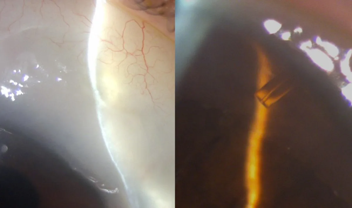
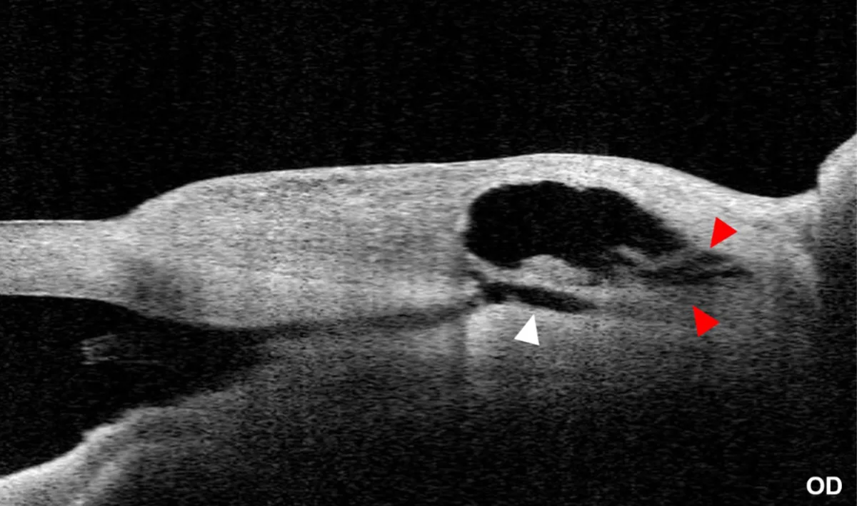
Right eye (OD) – Sub-Tenon’s XEN® Gel Stent implantation
Although the distal end of the device was not visible by the means of the slit-lamp examination, CASIA2 was able to confirm its sub-Tenon’s placement (white arrow head) and the presence of filtration at that level (red arrow heads).
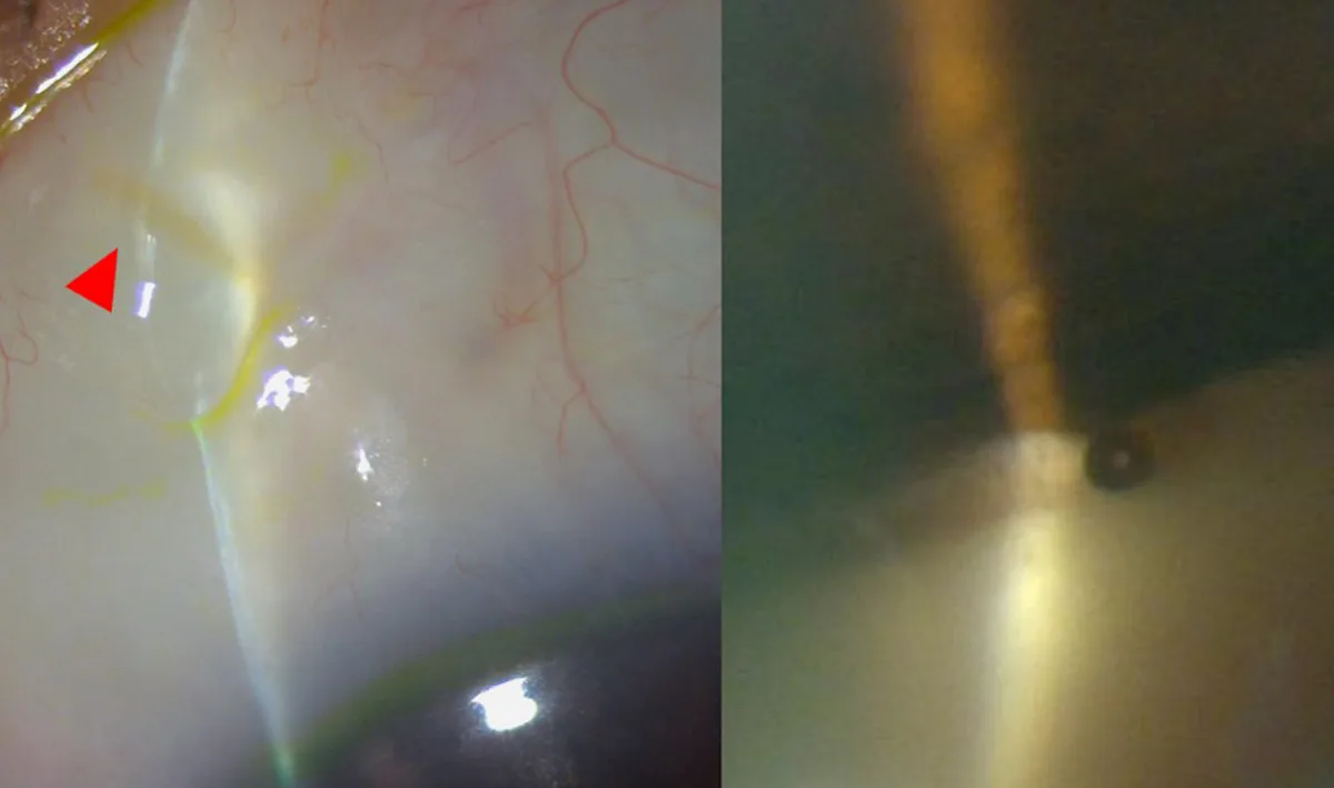
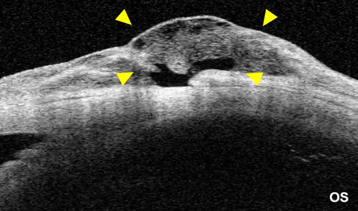
Left eye (OS) – Subconjunctival XEN® Gel Stent implantation
Positioning the distal end of the device (red arrow head) beneath the conjunctiva led to subconjunctival filtration (yellow arrow heads), and a hyporeflective bleb with microcysts. All of these features can be evaluated with CASIA2.
*AbbVie Inc., Chicago, Illinois, USA
Further Questions?
Do you have any questions or require further information?
We would be pleased if you send us an inquiry.