
Visualisation of Dual Intraocular Lenses Using CASIA2 Anterior Segment OCT
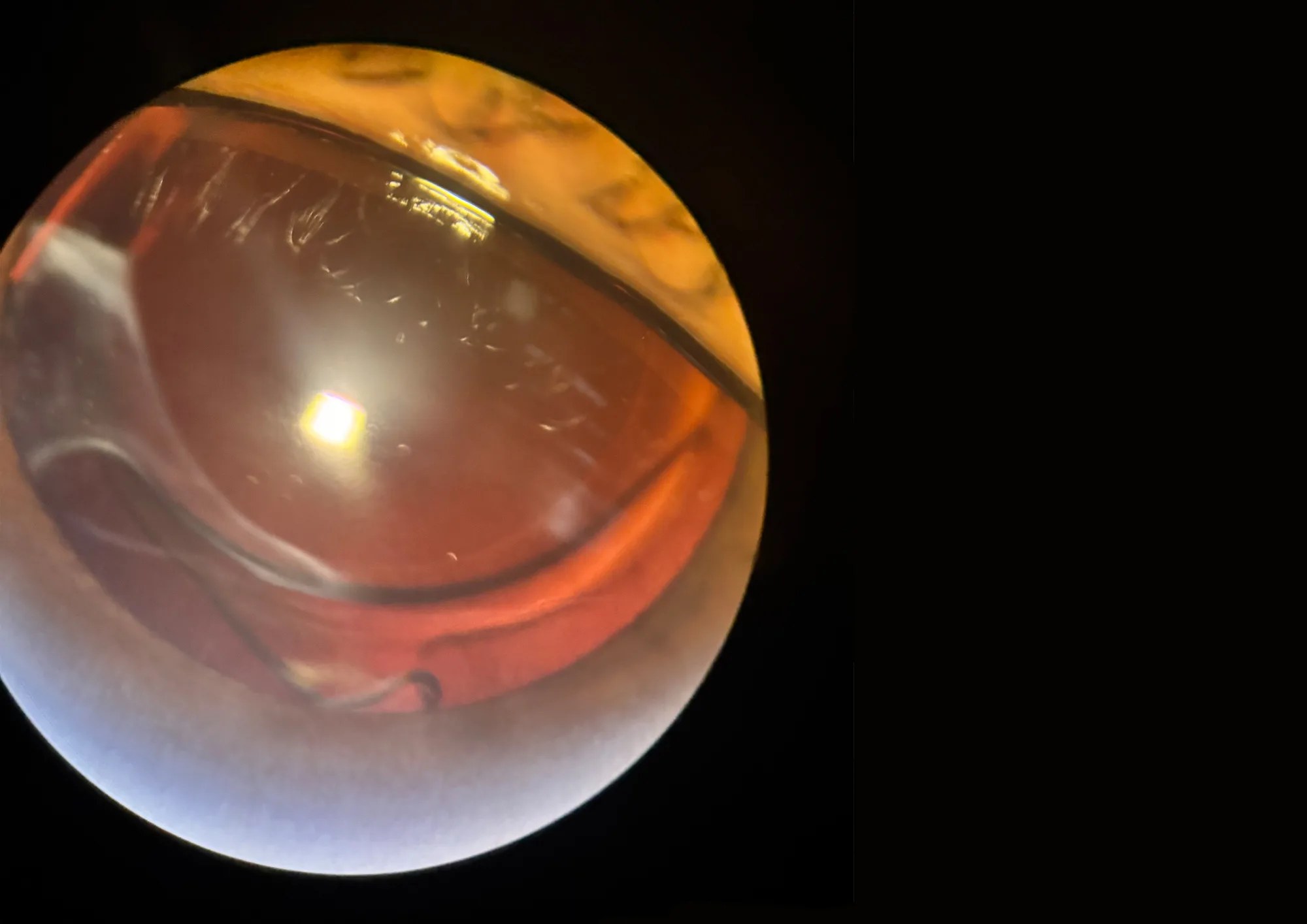
A patient with high myopia underwent refractive lens exchange with implantation of two intraocular lenses within the capsular bag. Dual intraocular lens implantation may be used in selected clinical cases to achieve the required refractive correction in patients with high myopia and extremely long axial length.
However, during routine slit-lamp examination, it is often difficult to clearly visualise the spatial configuration of two intraocular lenses located inside the capsular bag. To evaluate the anatomical structures of the anterior segment more precisely, imaging was performed using CASIA2 anterior segment OCT.
CASIA2 Examination
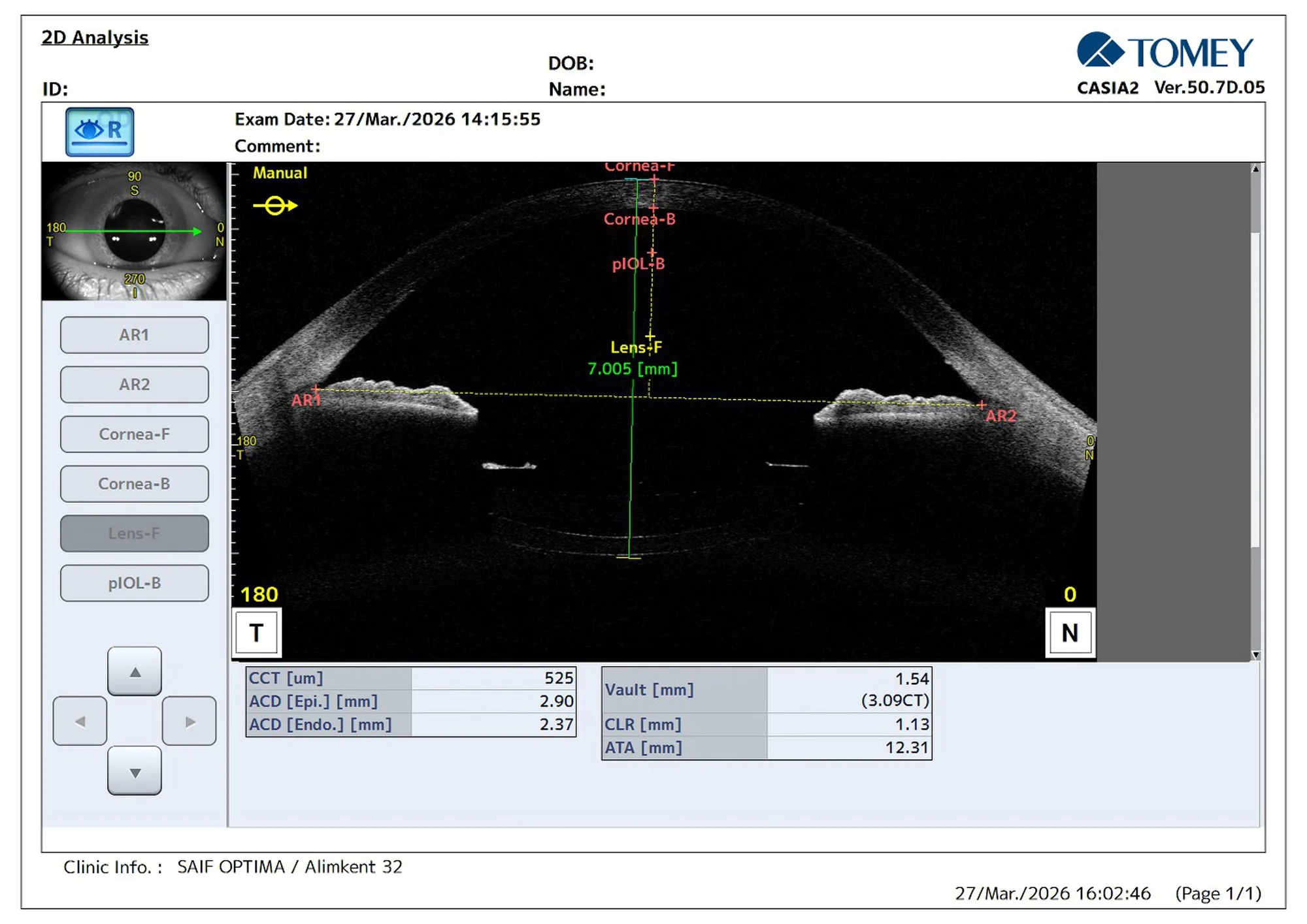
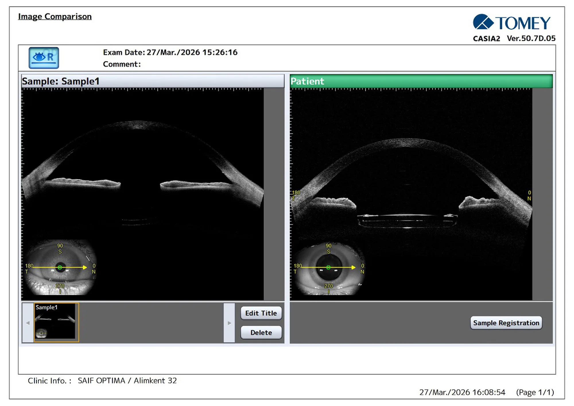
Anterior segment OCT imaging provided high-resolution cross-sectional images of the ocular structures.
The use of CASIA2 allowed clear visualisation of:
- the cornea
- the anterior chamber
- the capsular bag
- the position of the two intraocular lenses
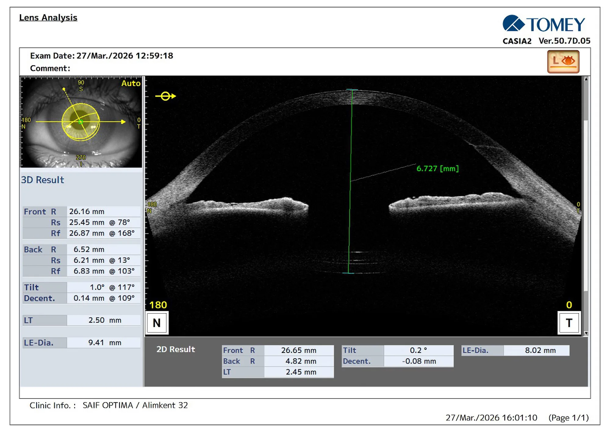
The OCT images clearly demonstrated the configuration of the two intraocular lenses positioned within the capsular bag.
Measurements
CASIA2 also allowed accurate measurements of important anatomical parameters of the anterior segment, including:
- anterior chamber depth
- distance from the cornea to the posterior capsule
- spatial configuration of the intraocular lens complex
These measurements provide objective information about the position of the lenses after surgery.
Discussion
Dual intraocular lens implantation can be an effective method for refractive correction in highly myopic eyes. However, evaluation of the spatial configuration of two intraocular lenses inside the capsular bag can be challenging using conventional slit-lamp examination. Anterior segment OCT imaging provides high-resolution cross-sectional visualisation of the anterior segment and allows precise assessment of intraocular lens position and configuration. Accurate visualisation of the intraocular lens complex is important for postoperative evaluation and for early detection of possible complications such as IOL decentration or interlenticular changes.
Clinical Value
Conventional diagnostic methods do not always allow precise visualisation of the spatial relationship between intraocular structures. The use of CASIA2 anterior segment OCT provides detailed imaging of the anterior segment and allows clear demonstration of the configuration of two intraocular lenses within the capsular bag. With this technology it is possible to measure the distance from the cornea to the posterior capsule and objectively evaluate the anatomical position of the intraocular lenses.
Educational Value
High-quality CASIA2 images are also extremely useful for scientific presentations and educational purposes. During scientific meetings it is not always possible to demonstrate surgical videos due to time limitations of presentations. Anterior segment OCT images allow surgeons to clearly demonstrate the position of intraocular lenses and explain the surgical concept quickly and effectively.
Conclusion
The use of CASIA2 anterior segment OCT allows precise visualisation of dual intraocular lenses within the capsular bag and enables accurate measurements of anterior segment anatomy. This technology represents a valuable diagnostic and educational tool for documenting the results of complex intraocular surgical procedures.
Acknowledgement
The author expresses sincere gratitude to her late mentor Ikbol Fazilovich Saliev, who first developed and patented the surgical concept of dual intraocular lens implantation. His innovative ideas laid the foundation for the surgical approach presented in this report.
1 / 4
2 / 4
Further Questions?
Do you have any questions or require further information?
We would be pleased if you send us an inquiry.