
Post-Surgery Corneal Edema and Athalamia in Glaucoma
A 45-year-old woman underwent phakic anterior chamber lens surgery 23 years ago. She develops glaucoma and undergoes a trabeculectomy. The day after surgery the chamber is wide, the IOP is 5 mmHg.
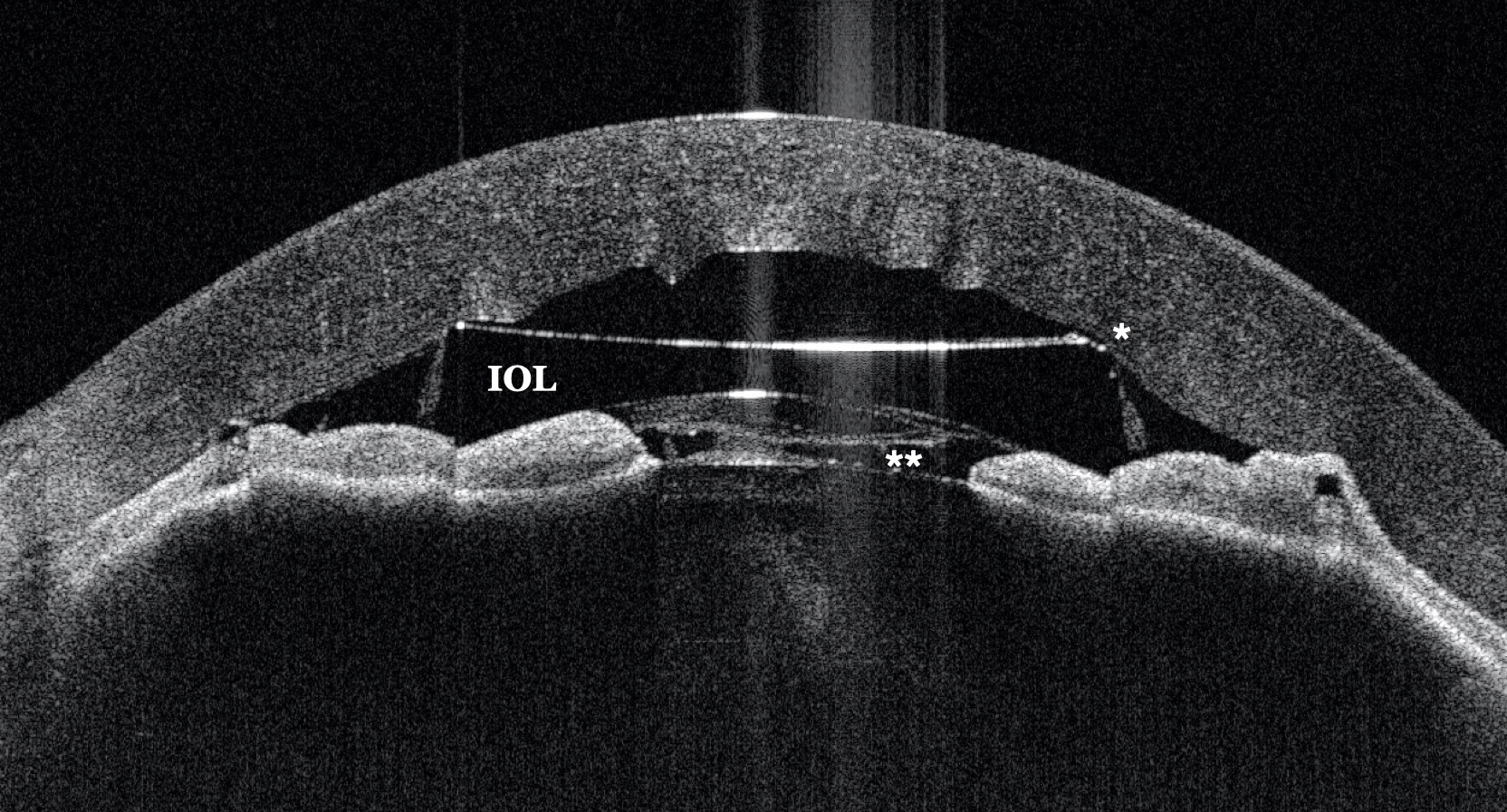
At the weekly check-up, the patient presented with corneal edema and athalamia (Figure 1). She underwent intensive treatment with mydriatic agents and corticosteroids. Performing an AS-OCT using CASIA2 allows all parameters to be monitored qualitatively and quantitatively: corneal edema, distance from the lens to the endothelium(*), irido-corneal contact, and the inflammatory pupillary membrane(**).
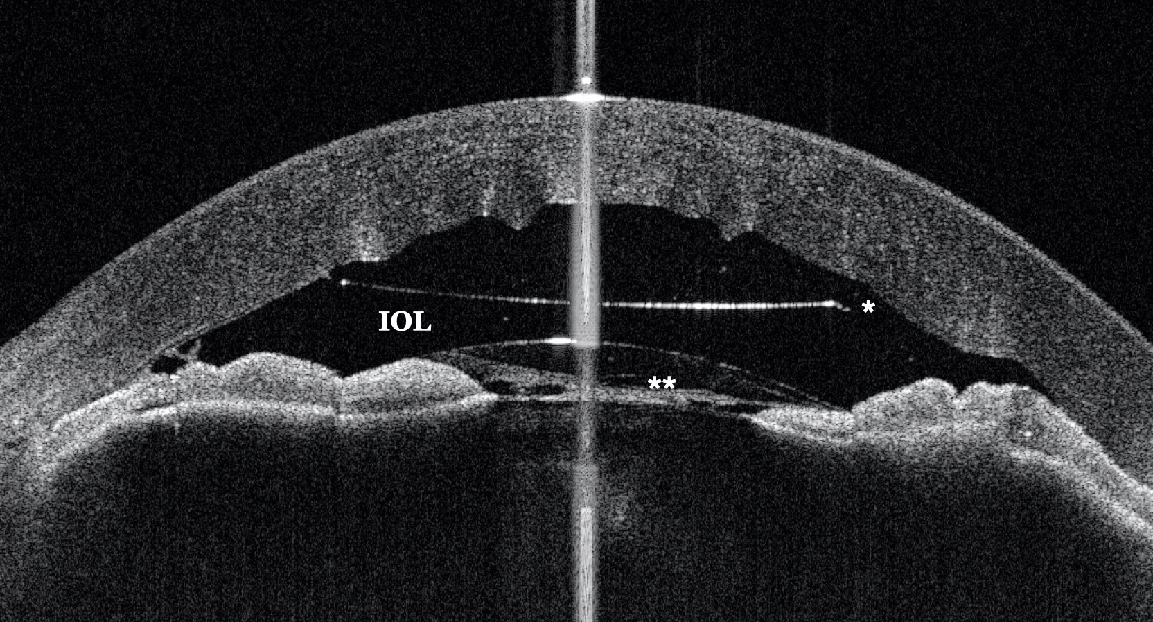
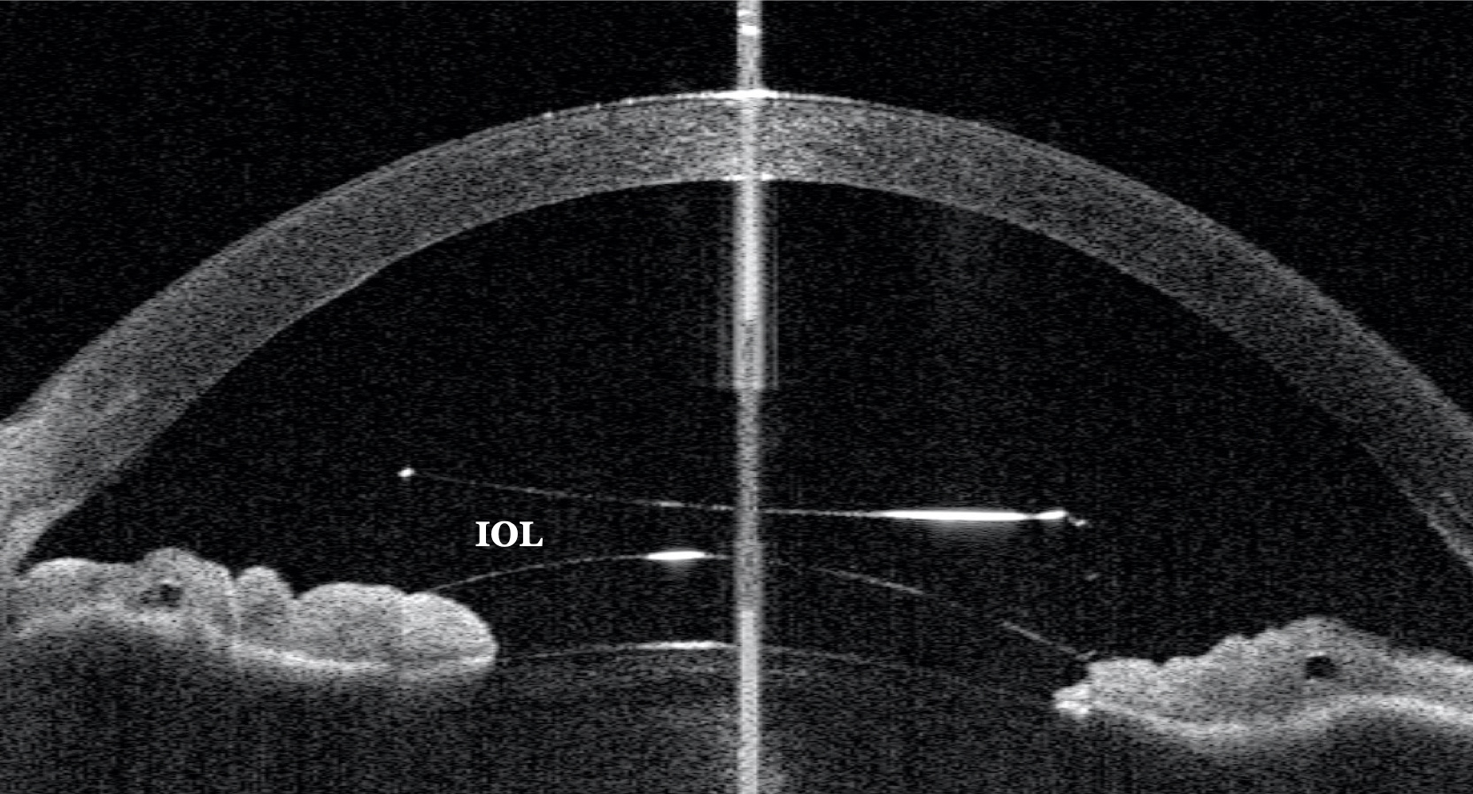
After 30 minutes the improvement was already visible on the OCT (Figure 2), and the next day the improvement could be quantified. In 3 days the clinical condition was resolved (Figure 3).
Further Questions?
Do you have any questions or require further information?
We would be pleased if you send us an inquiry.