
Pellucid Marginal Degeneration
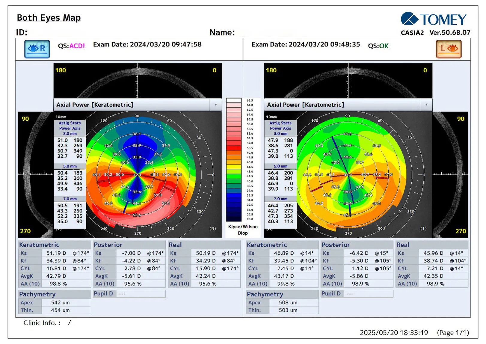
A 74-year-old female patient presented with gradually decreasing vision, particularly in her right eye, with notable difficulties at night and an increase in astigmatism that her regular glasses no longer corrected adequately. The best corrected visual acuity (BCVA) in the right eye (OD) was 0.2 with high against-the-rule astigmatism, approximately -12.00D × 110°, and in the left eye (OS) it was 0.5 with moderate against-the-rule astigmatism.
Topographic examination revealed a classic “crab claw” pattern on axial power maps, particularly in the right eye, with marked inferior corneal steepening and flattening in the vertical meridian above the thinned area. Against-the-rule astigmatism was evident, with the right eye showing a steep K (Ks) of approximately 51D and a flat K (Kf) of around 35 D, resulting in significant corneal astigmatism (>15 D).
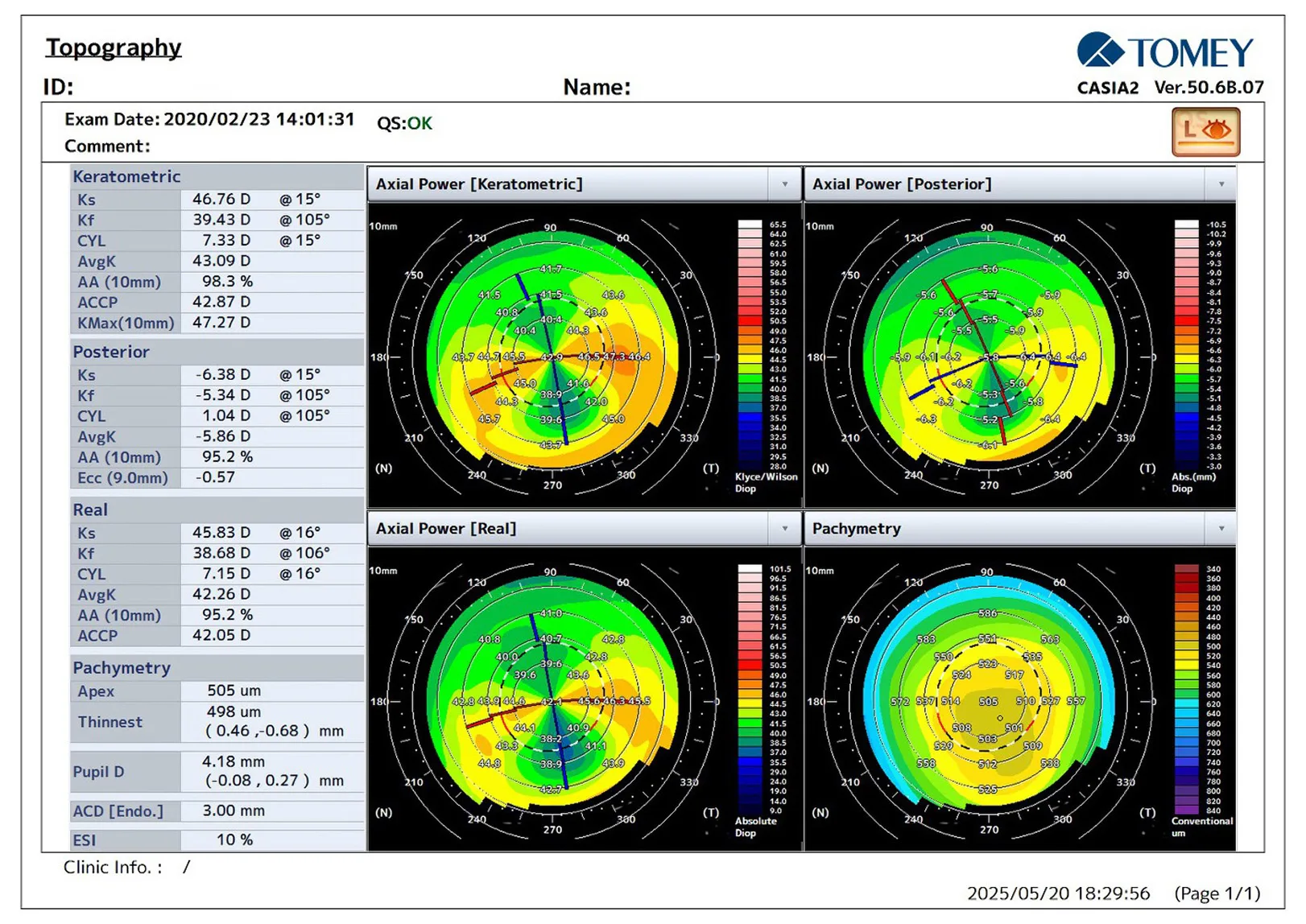
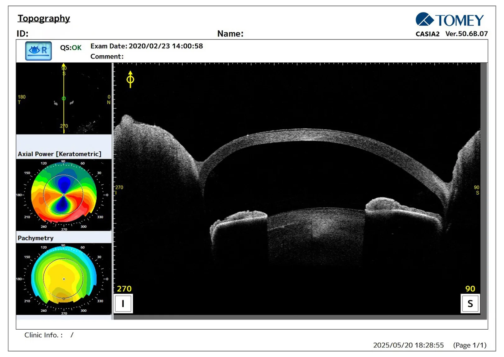
In the left eye, the astigmatism was less pronounced but still significant (6-8 D). The minimum corneal thickness was approximately 450 μm in the right eye and 500 μm in the left eye. Vertical scans displayed normal central corneal architecture with abrupt thinning in the inferior peripheral cornea. CASIA2 imaging confirmed the diagnosis of pellucid marginal degeneration rather than inferior keratoconus, characterised by peripheral rather than paracentral thinning and a distinctive crab claw topographic pattern.
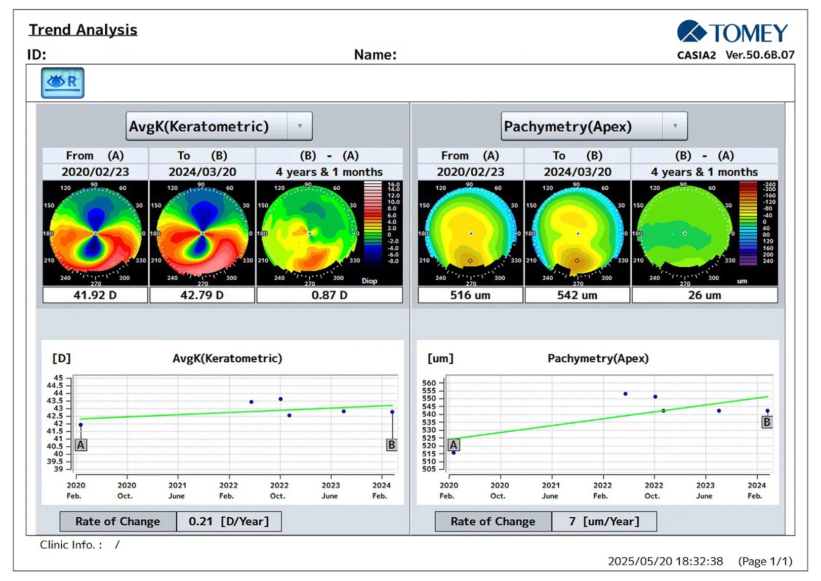
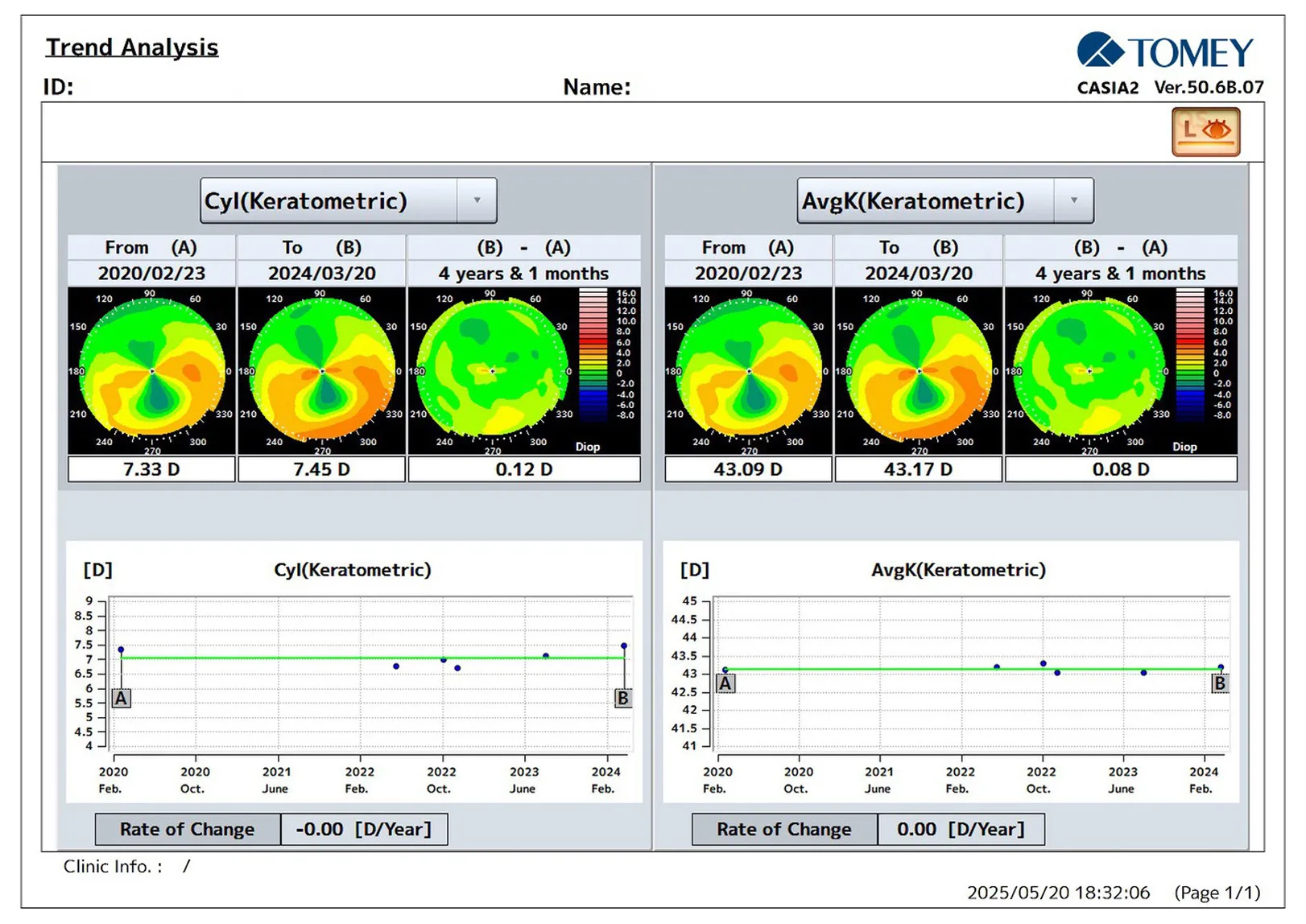
This trend analysis compares keratometric data over a span of 4 years and 1 month. The average keratometric power has remained consistent, with minimal variation of 0.87 D in the right and 0.08 D in the left eye.
Further Questions?
Do you have any questions or require further information?
We would be pleased if you send us an inquiry.