
Management of Capsular Bag Phimosis
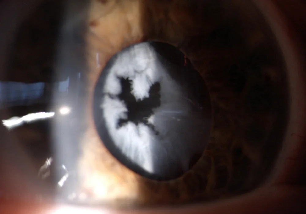
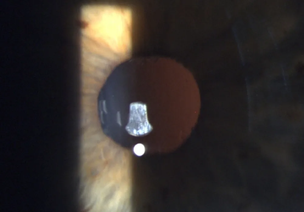
A 63-year-old woman was referred for laser capsulotomy for capsular phimosis diagnosed three months after uneventful bilateral cataract surgery. During examination, pinhole capsular phimosis was noted.
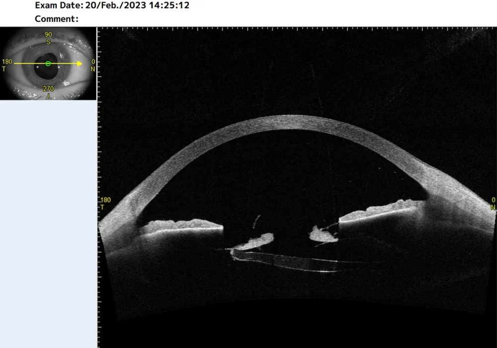
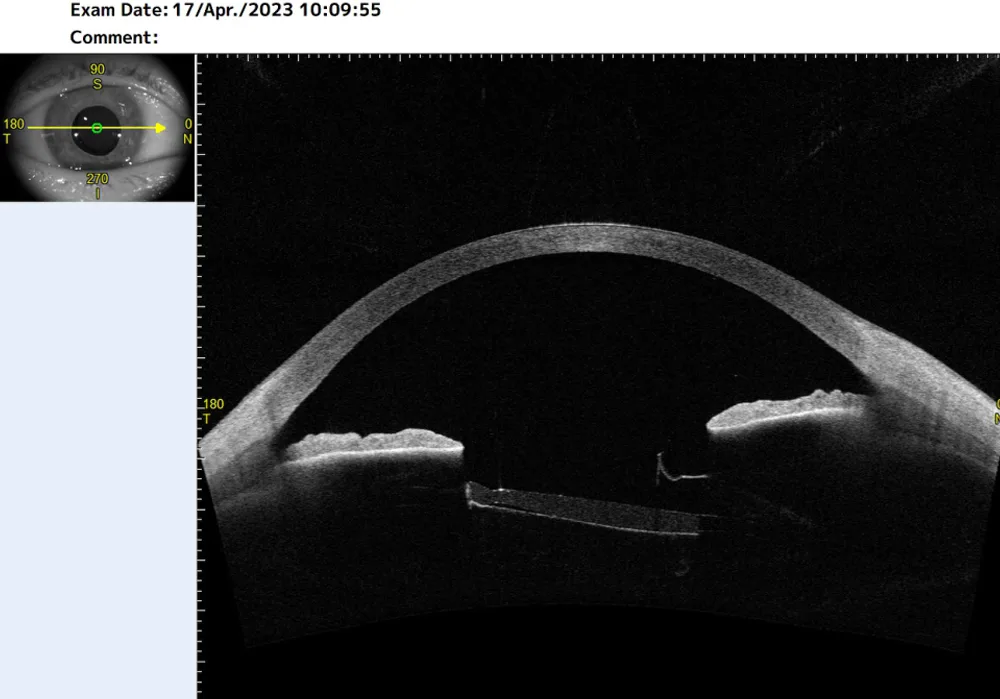
Anterior segment OCT imaging was employed to document and visualise the extent and unusual thickness of the phimotic capsular bag. Nd:YAG laser capsulotomy was performed over three sessions, with 150-600 mJ of laser energy applied per eye per session. OCT was utilised to monitor the progress of the interrupted surgical treatment, allowing for documentation of the capsulotomy’s extent and effectiveness (Figure 1: after first capsulotomy, Figure 2: follow-up visit after final treatment).
For postoperative treatment, NSAID and cortisone eye drops were prescribed. Despite this, the patient developed mild cystoid macular edema postoperatively. With continued treatment, macular edema resolved over a one-month period.
OCT was utilised to document the extent and unusual thickness of the phimotic capsular bag and allowed for assessment of the progress during the multi-session treatment.
Further Questions?
Do you have any questions or require further information?
We would be pleased if you send us an inquiry.