
Macular Corneal Dystrophy
A 45-year-old female patient with a significant family history of macular dystrophy presented with complaints of reduced vision. Her best corrected visual acuity (BCVA) was 0.8 in the right eye (OD) and 0.2 in the left eye (OS).
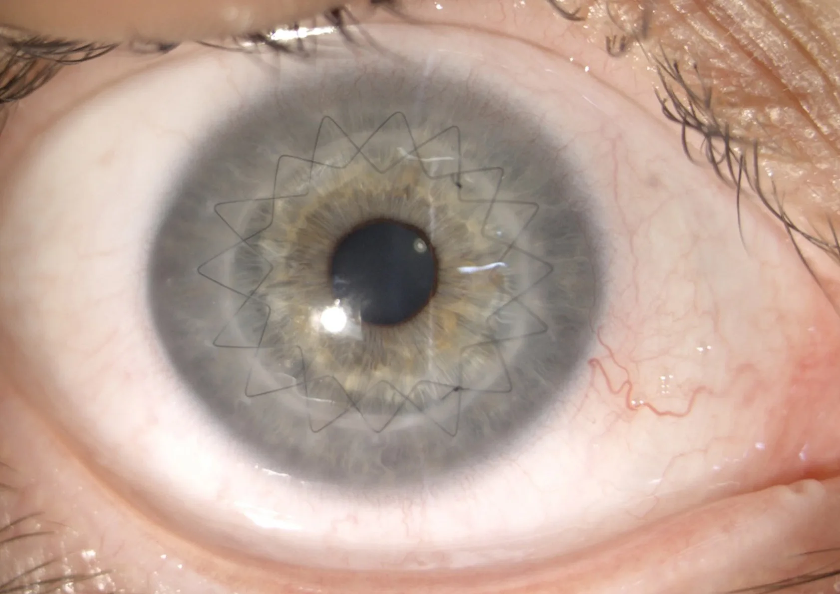
The right eye had undergone penetrating keratoplasty (PKP) 7 months prior due to corneal pathology. The left eye had no surgical history but presented with progressive visual deterioration.
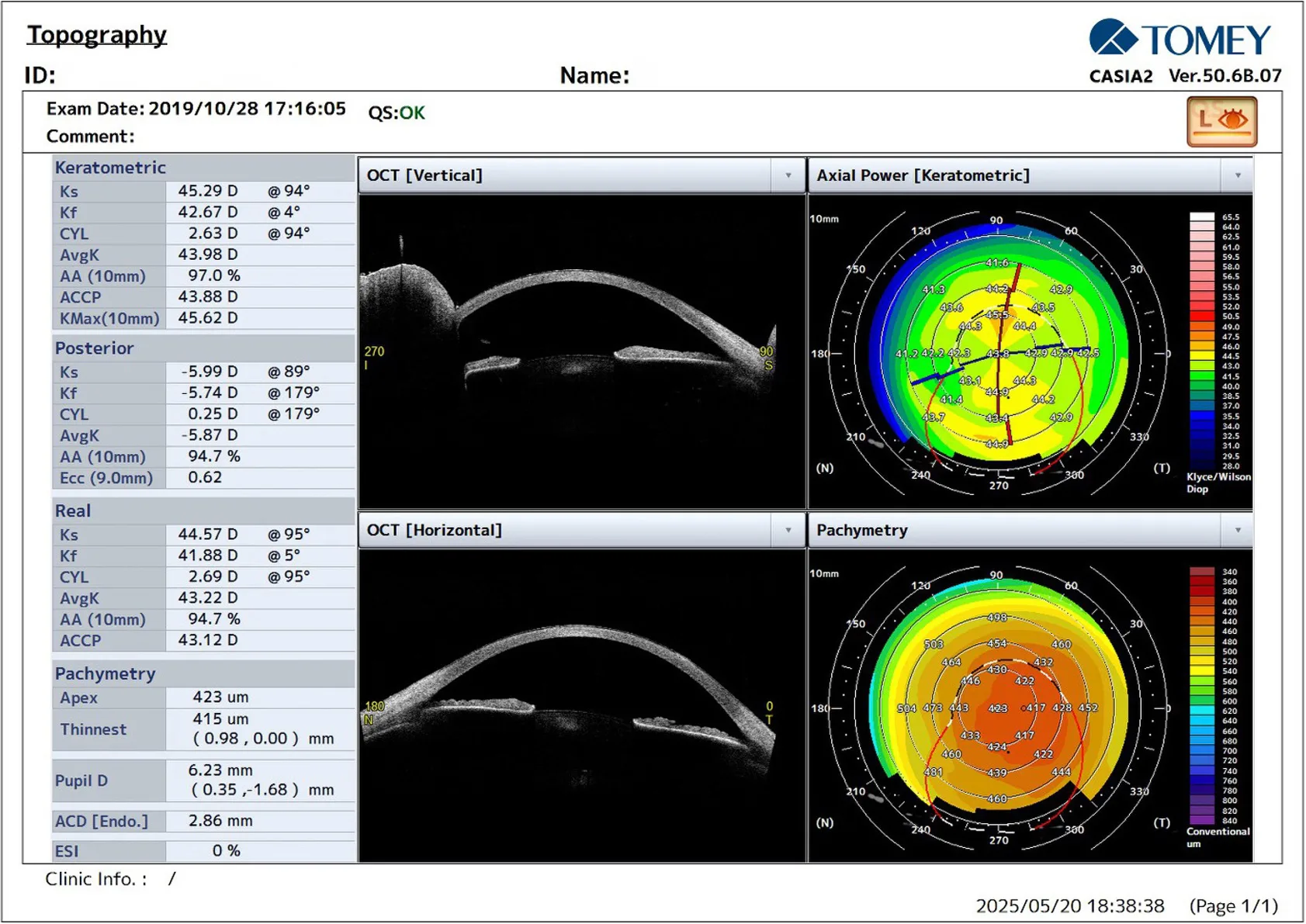
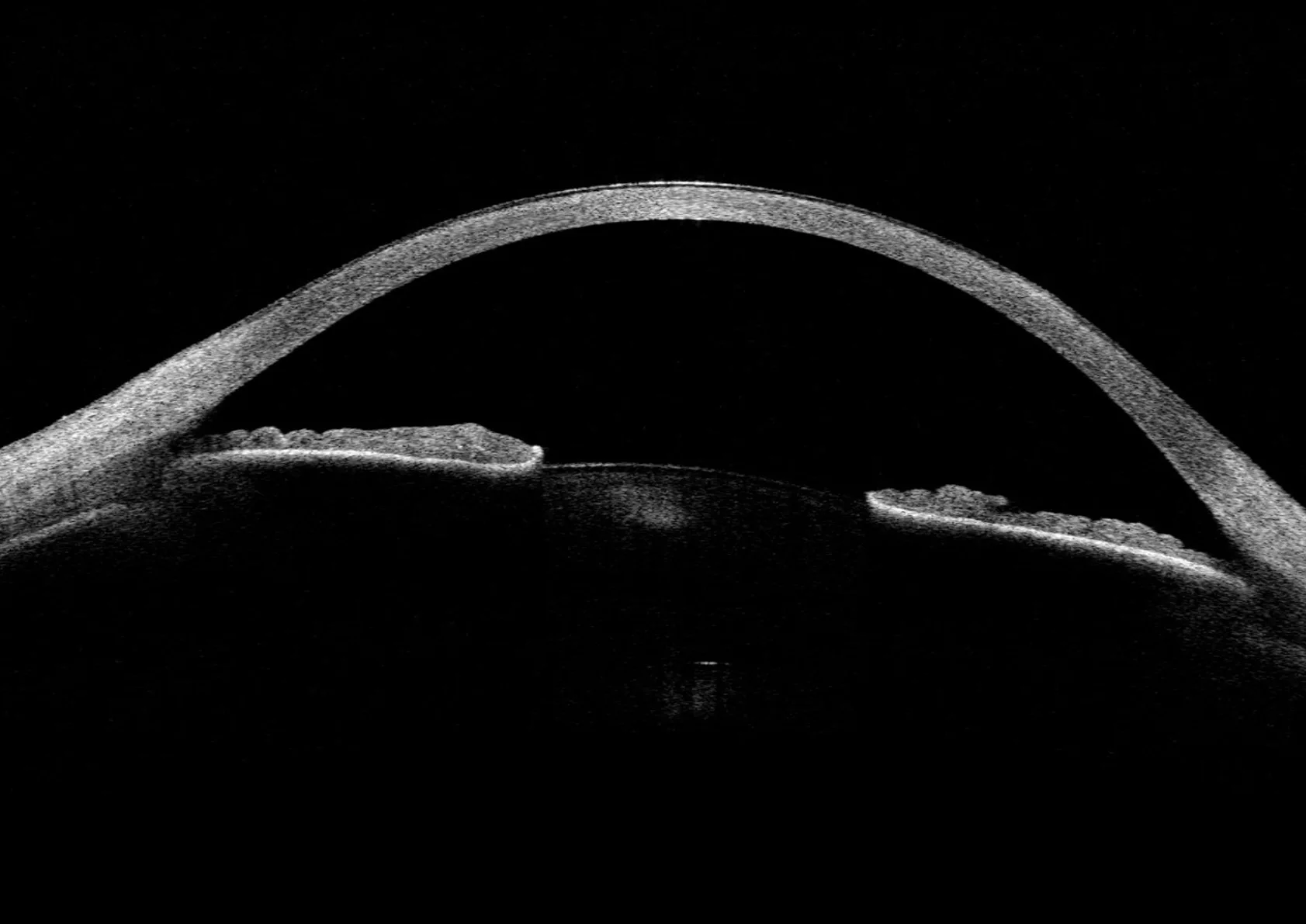
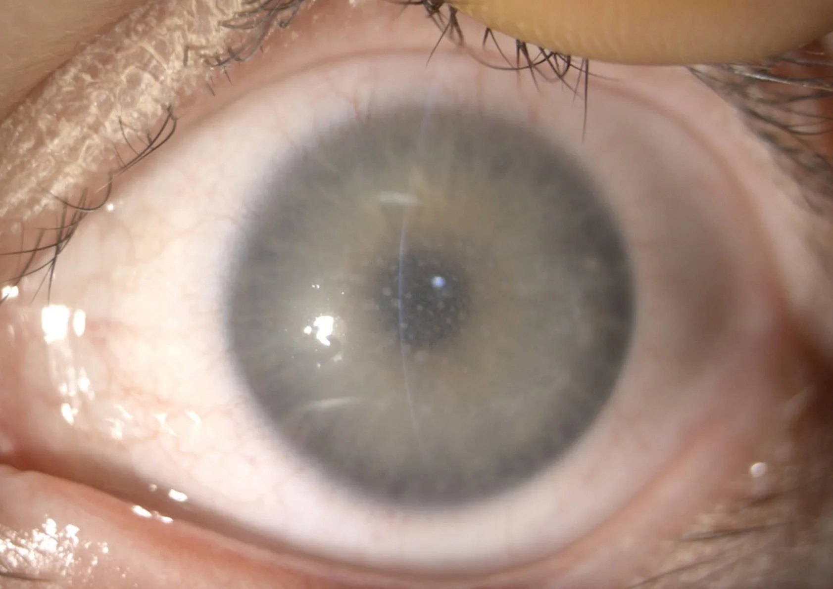
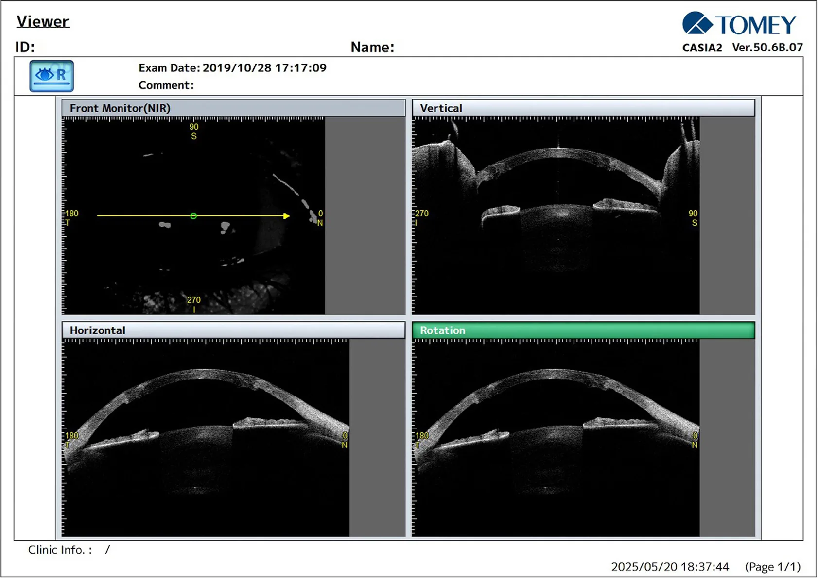
The left eye showed generalised corneal thinning and the increased corneal stromal reflectivity characteristic of macular dystrophy.
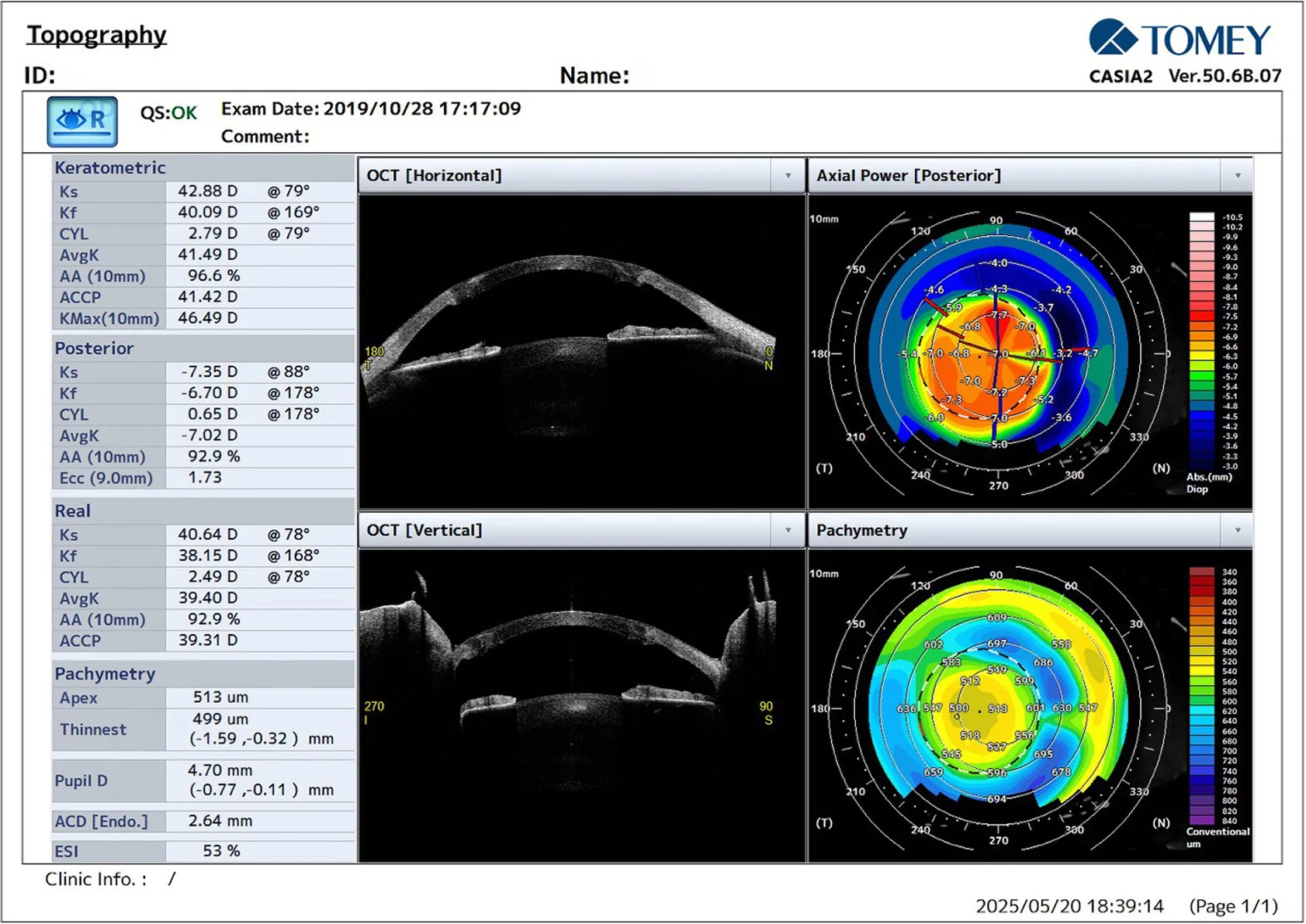
The graft appeared structurally intact with acceptable thickness centrally and regular posterior corneal contour. AvgK post-PKP was 41.49 D, with residual astigmatism of 2.79 D. The CASIA2 anterior segment OCT proved crucial in evaluating corneal structure and detecting macular corneal dystrophy, particularly in the left eye. The right eye showed a stable post-PKP graft with residual regular astigmatism.
Further Questions?
Do you have any questions or require further information?
We would be pleased if you send us an inquiry.