
Iatrogenic Injury of the Lens Capsule
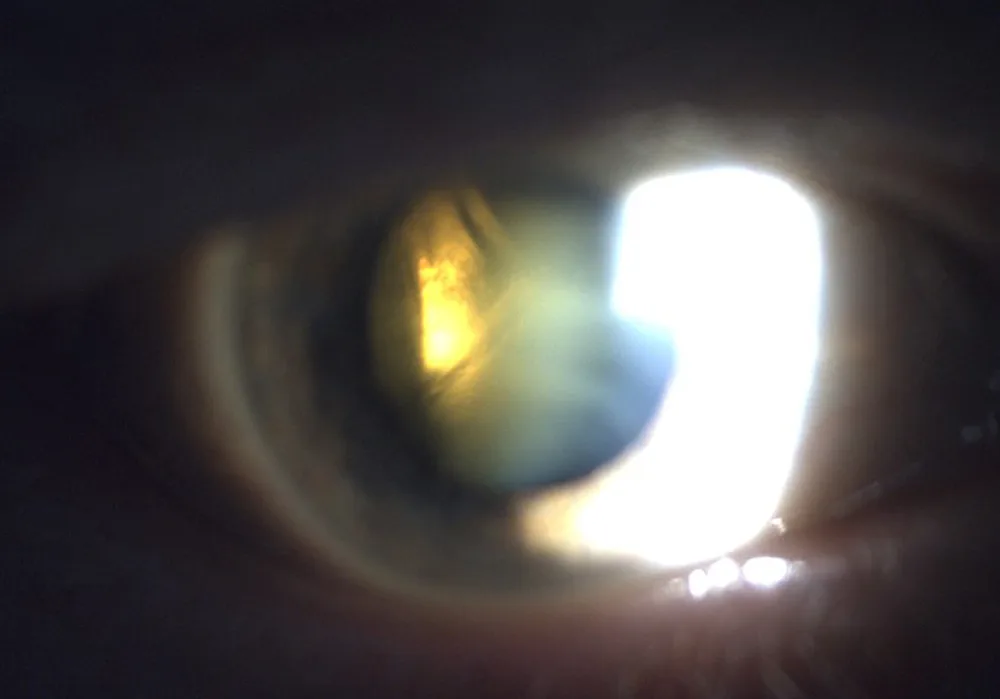
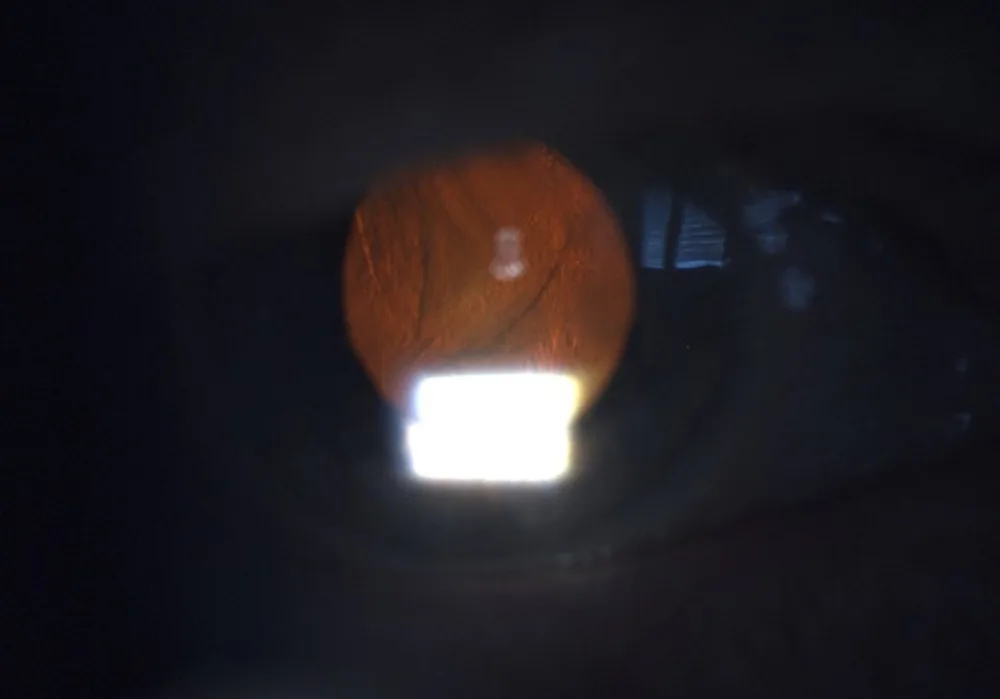
An 80-year-old woman presented with worsening vision five days after an intravitreal anti-vascular endothelial growth factor injection for age-related macular neovascularisation. The patient’s file noted early dementia and reduced cooperation during intravitreal injections. Best-corrected visual acuity (BCVA) had declined from 0.5 pre-injection to 0.05 at presentation. Slit-lamp examination revealed a clear cornea, quiet anterior chamber, nuclear cataract, and probable defect of the posterior lens capsule (Figures 1 and 2). The fundus view was limited, but no signs of endophthalmitis were recorded.
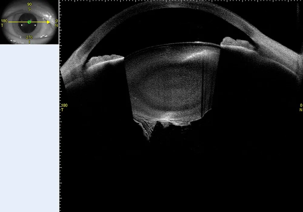
Anterior segment OCT demonstrated a posterior lens capsule injury (Figure 3), suspected to be iatrogenic from the recent intravitreal injection. This diagnosis guided the decision to perform lens extraction under general anesthesia with vitrectomy standby.
Intraoperatively, the capsular defect led to nucleus drop into the vitreous cavity, necessitating vitrectomy and sulcus intraocular lens implantation. Postoperatively, BCVA improved to 0.63 after nine days.
This case highlights the utility of high-resolution anterior segment OCT in definitively diagnosing posterior capsule injuries. The imaging modality provided objective preoperative information, allowing for appropriate surgical planning in managing this uncommon complication of intravitreal injections.
Further Questions?
Do you have any questions or require further information?
We would be pleased if you send us an inquiry.