
Descemet’s Detachment – Rare Post-NPGS Complication
A 65-year-old patient underwent deep sclerectomy – non-penetrating glaucoma surgery (NPGS) – for glaucoma five years ago. The IOP was maintained at levels between 11-13 mmHg during this time. He went to the emergency room due to decreased visual acuity in the operated eye. IOP was 12 mmHg.
Corneal edema was observed and treatment was started with topical corticosteroids and hyperosmolar agents. He was referred to the Glaucoma Unit.
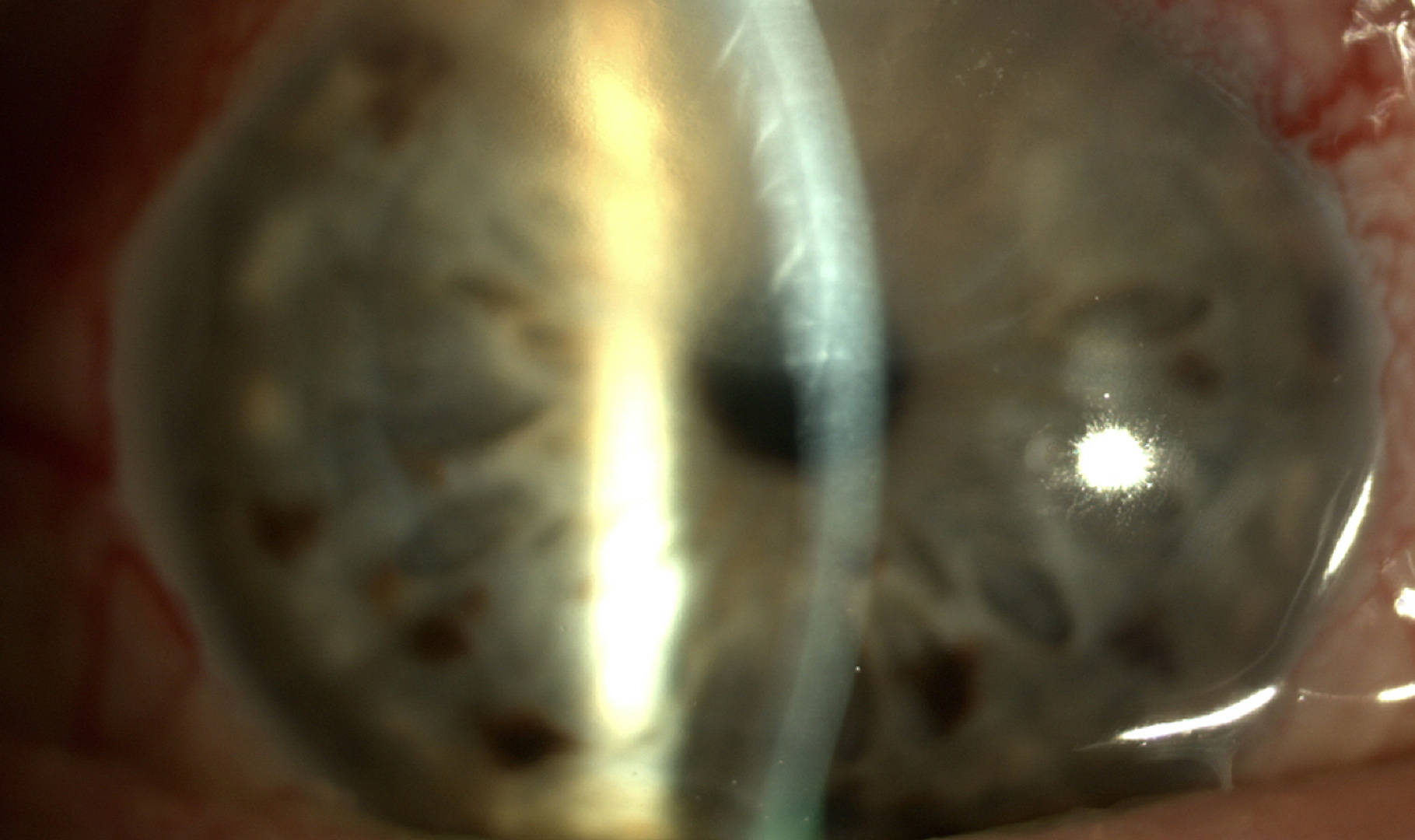
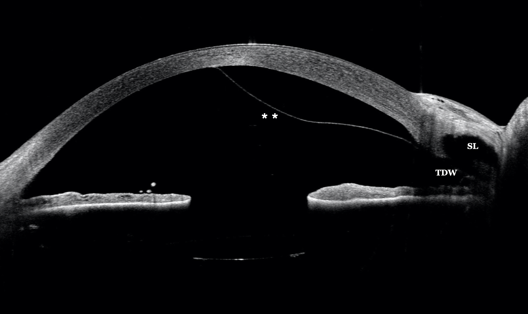
On examination we found a superior, semicircular, and well-differentiated corneal edema (Figure 1). For the correct diagnosis, AS-OCT is performed using CASIA2.
In the surgical area, the scleral lake (SL) and the trabeculo-Descemet window (TDW) can be seen. There is corneal thickening and detachment of Descemet’s membrane (**) (Figure 2).
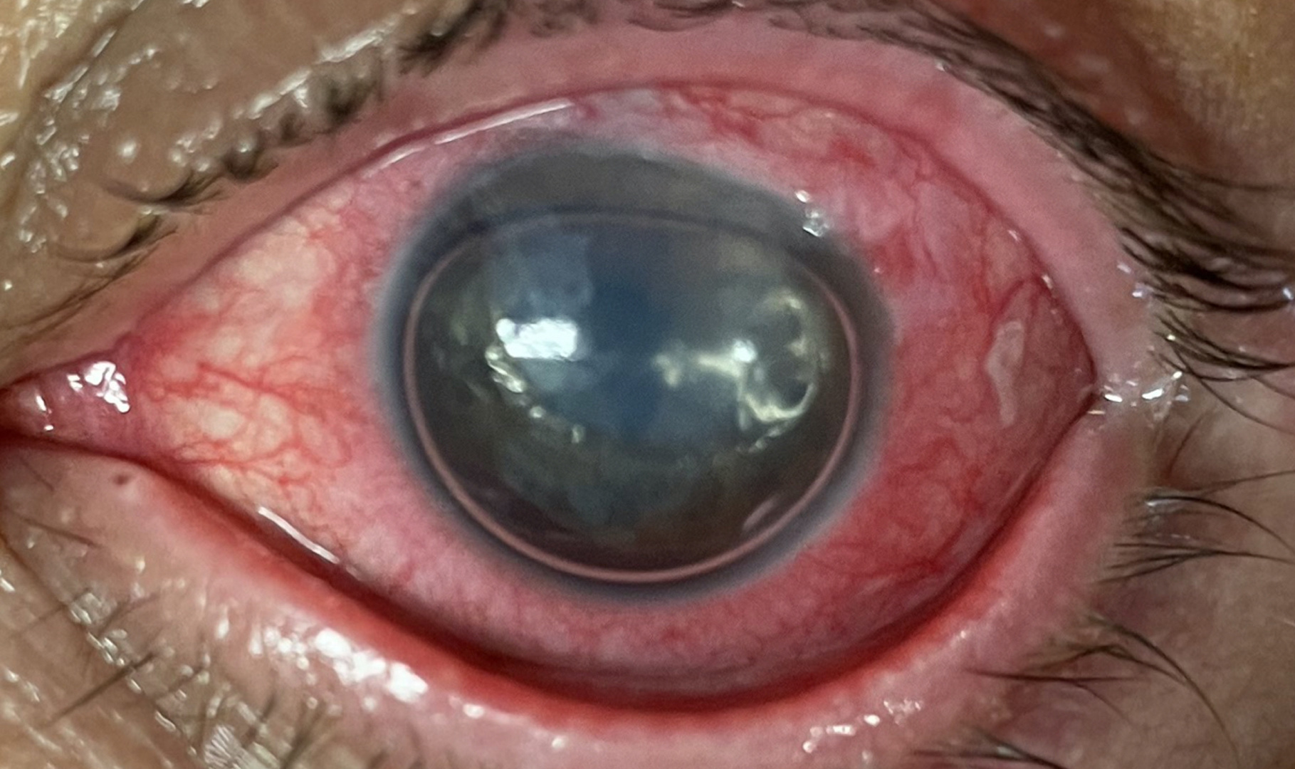
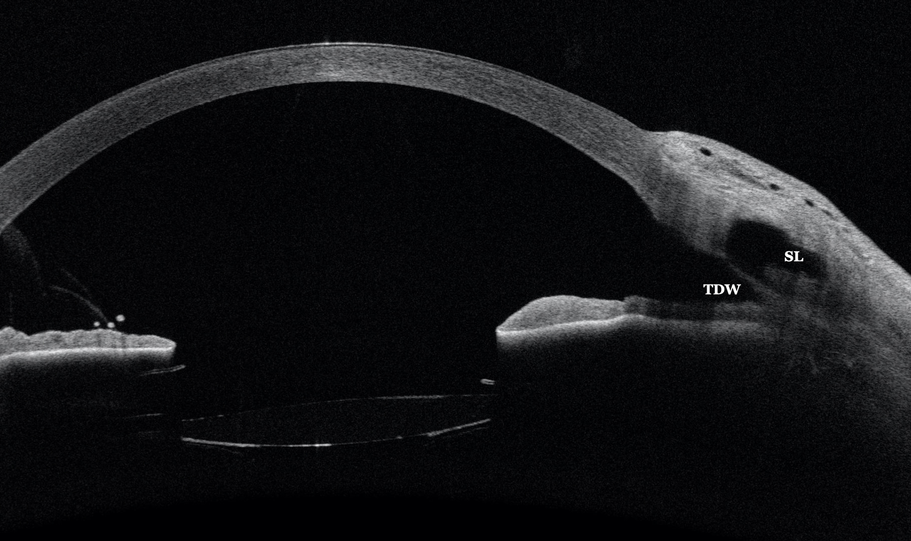
Descemet’s membrane detachment is an unusual complication of NPGS that is often associated with an increase in IOP due to a tenon cyst. On this occasion the IOP was within normal limits. Its diagnosis is very complex if AS-OCT is not available, because with the slit lamp it is not possible to differentiate the Descemet’s membrane due to the clouding of the cornea. The treatment consists of injection of 20% SF6 in the anterior chamber (Figure 3) which manages to apply the membrane. This is constable using the AS-OCT (Figure 4).
Further Questions?
Do you have any questions or require further information?
We would be pleased if you send us an inquiry.