
Post-ACXL Corneal Edema in Advanced Keratoconus
Patient: 27-year-old male
Diagnosis: Bilateral keratoconus — OD: stage III, OS: stage IV
Procedure: Accelerated corneal cross-linking (ACXL), Epi-Off (9 mW/cm2, 10 minutes) on OD
Clinical Context
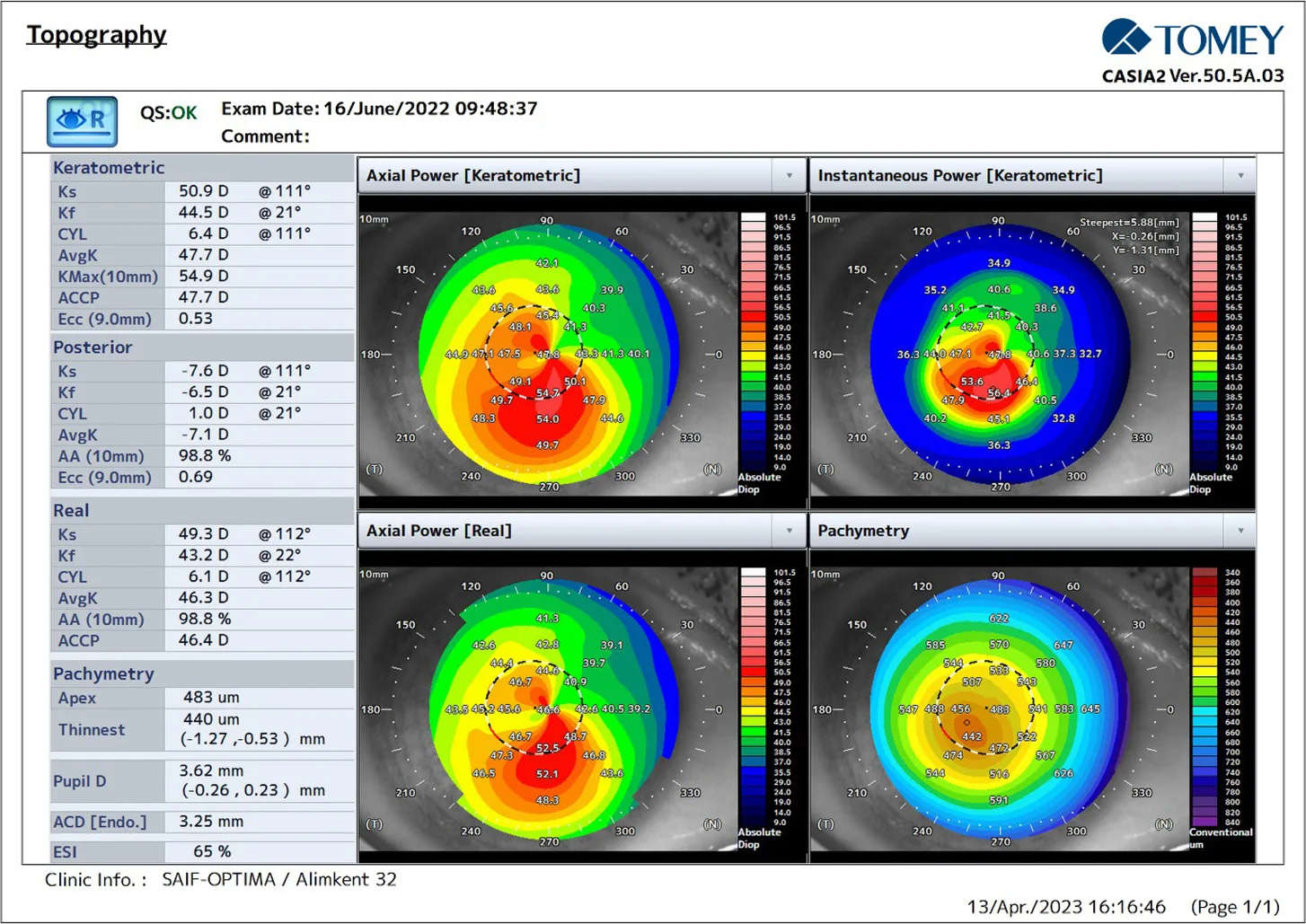
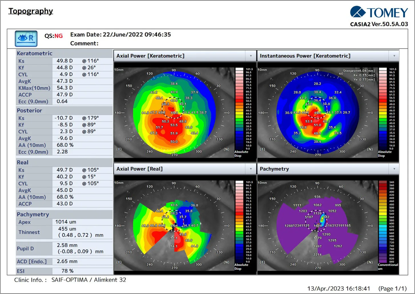
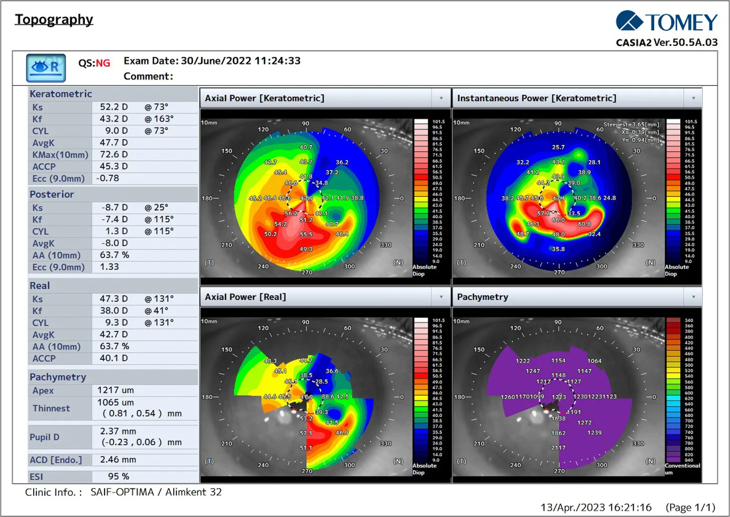
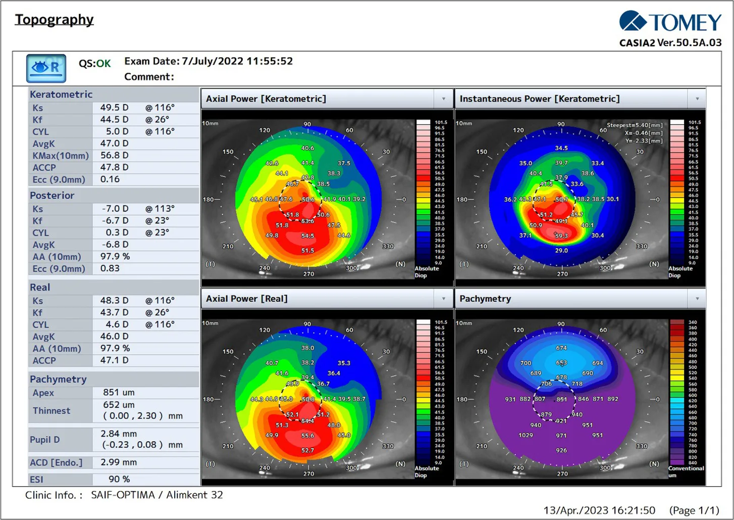
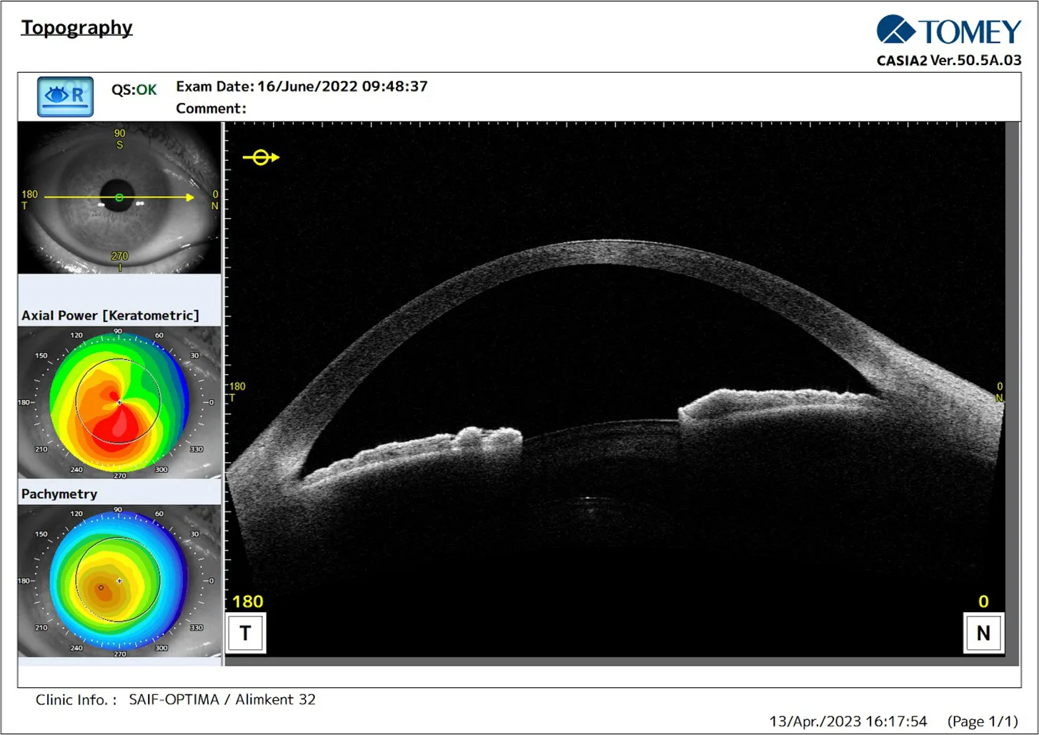
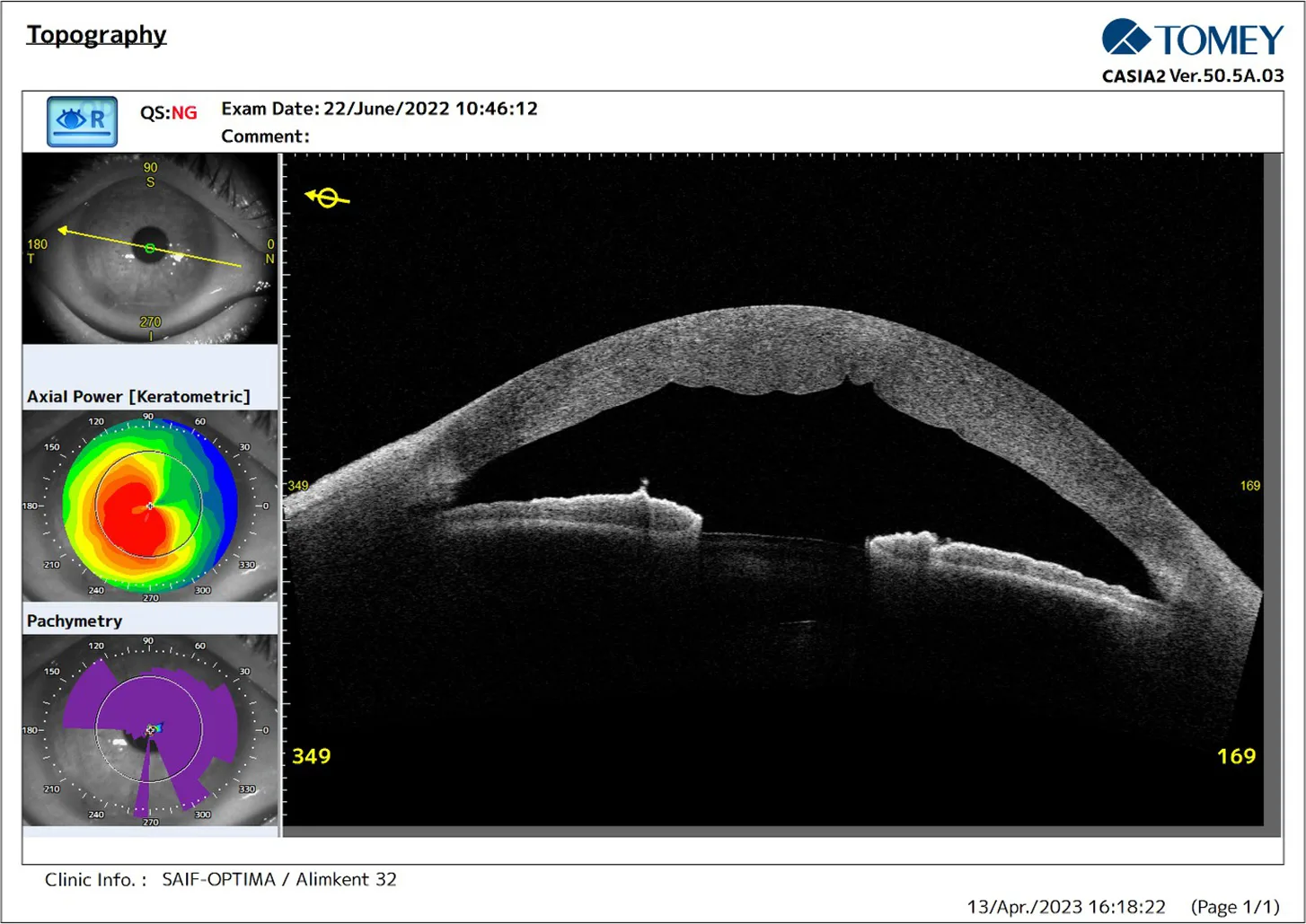
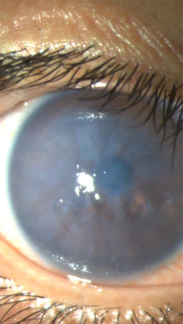
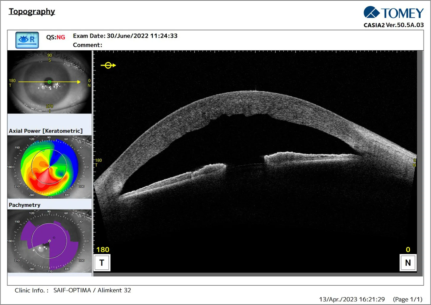
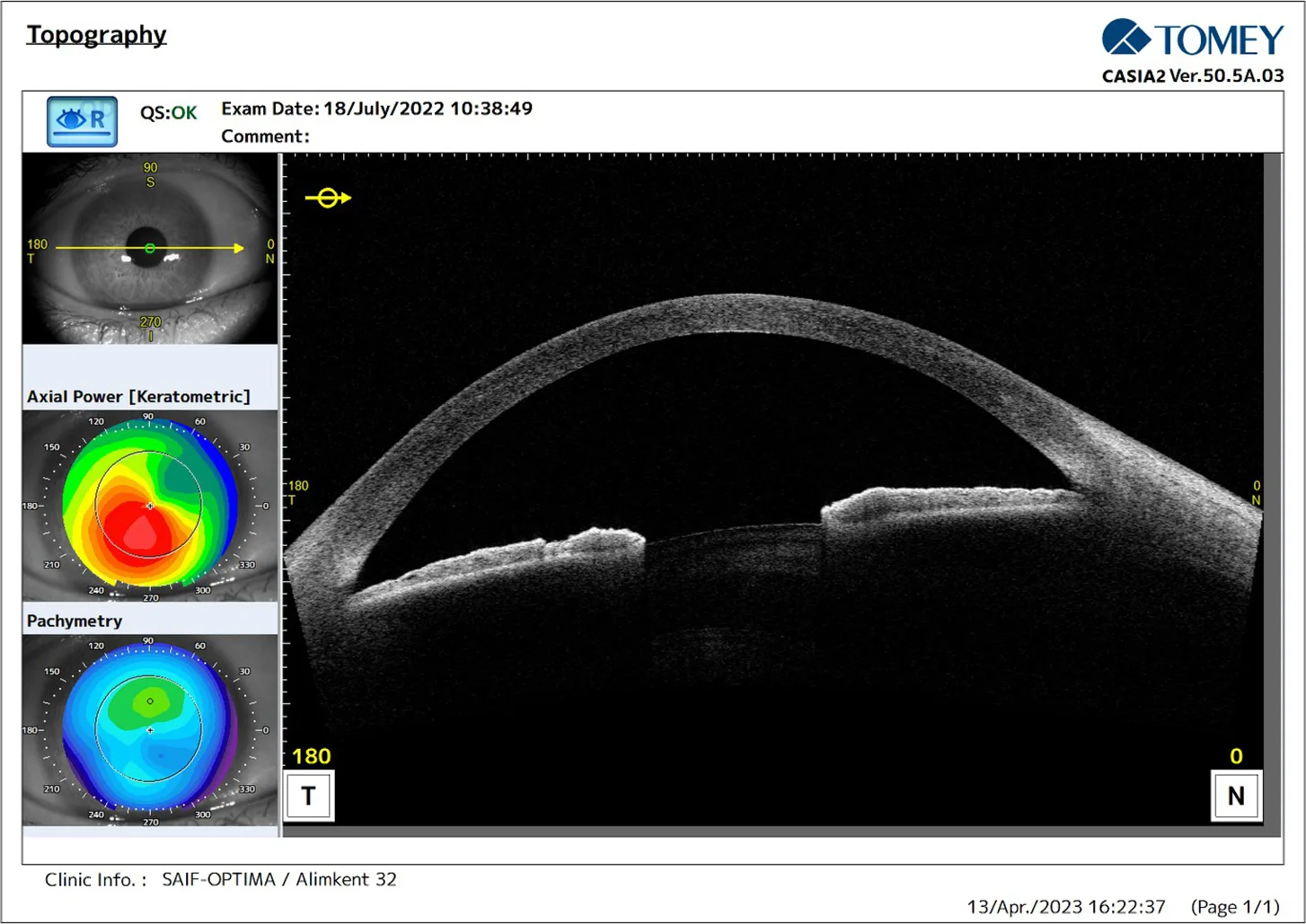
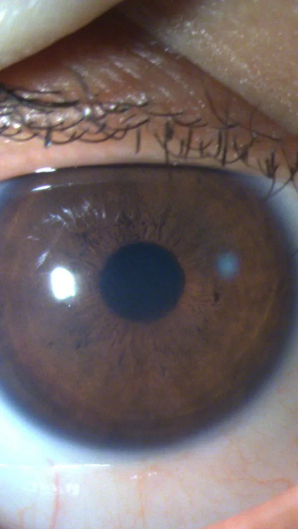
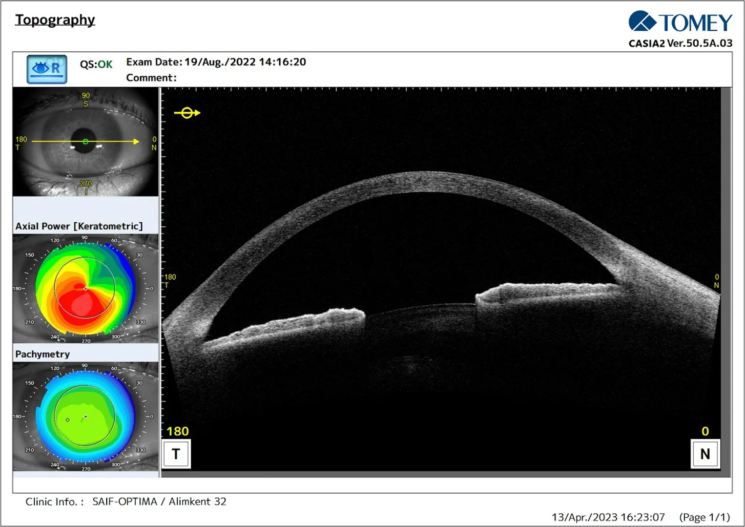
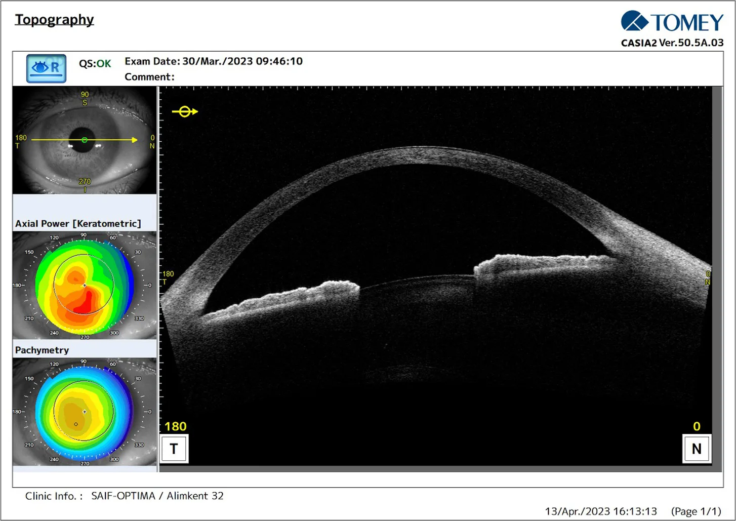
Following ACXL, the patient presented with visible stromal edema and Descemet’s membrane folds. Slit-lamp visualisation was limited by corneal haze. The need for accurate, reproducible, and non-invasive imaging was clear. CASIA2 provided the precision and depth of information necessary to track the healing process over time.
Key Contributions of CASIA2 in this Case
- Enabled detailed layer-by-layer imaging, even in the presence of haze
- Provided objective validation of natural recovery
- Helped avoid unnecessary treatment through precise monitoring
- Ensured safe long-term follow-up with clarity and consistency
Conclusion: More Than a Device – A New Standard of Care
The CASIA2 is not just an imaging device – it is a revolution in anterior segment diagnostics. In this case, it offered what no slit lamp or ultrasound could: a non-invasive, highly detailed, and dynamically trackable view of the cornea’s healing journey.
Its ability to visualise microstructural changes with such depth and accuracy makes it indispensable in modern ophthalmology. For corneal surgeons, refractive specialists, and glaucoma experts alike, CASIA2 raises the standard, shortens decision-making time, and enhances patient outcomes.
Once you start working with CASIA2, you will no longer imagine practicing without it. It is more than a tool – it is a clinical ally, a precision instrument, and for many of us it is the eye behind our eyes.
Why CASIA2?
The CASIA2 swept-source OCT is a transformative advance in anterior segment imaging. With its unparalleled resolution, deep scanning depth, and ultra-fast acquisition, it offers a level of clarity, confidence, and control that is simply unmatched.
Whether it’s for keratoconus diagnostics, angle assessment, ICL planning, or post-op monitoring, CASIA2 gives clinicians the tools they need to see more, understand in more depth, and act sooner.
| CASIA2 Findings | Clinical Relevance | |
|---|---|---|
| Day 1 | Stromal edema, Descemet’s folds | Initial post-op response |
| Day 7 | Edema unchanged | Safe observation confirmed |
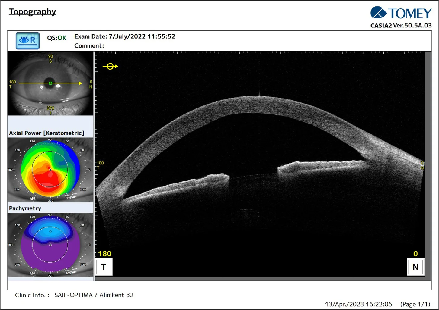
| Day 14 | Folds resolved, edema decreasing | Signs of spontaneous recovery |
| Day 30 | Clear cornea, regular epithelium | Clinical resolution |
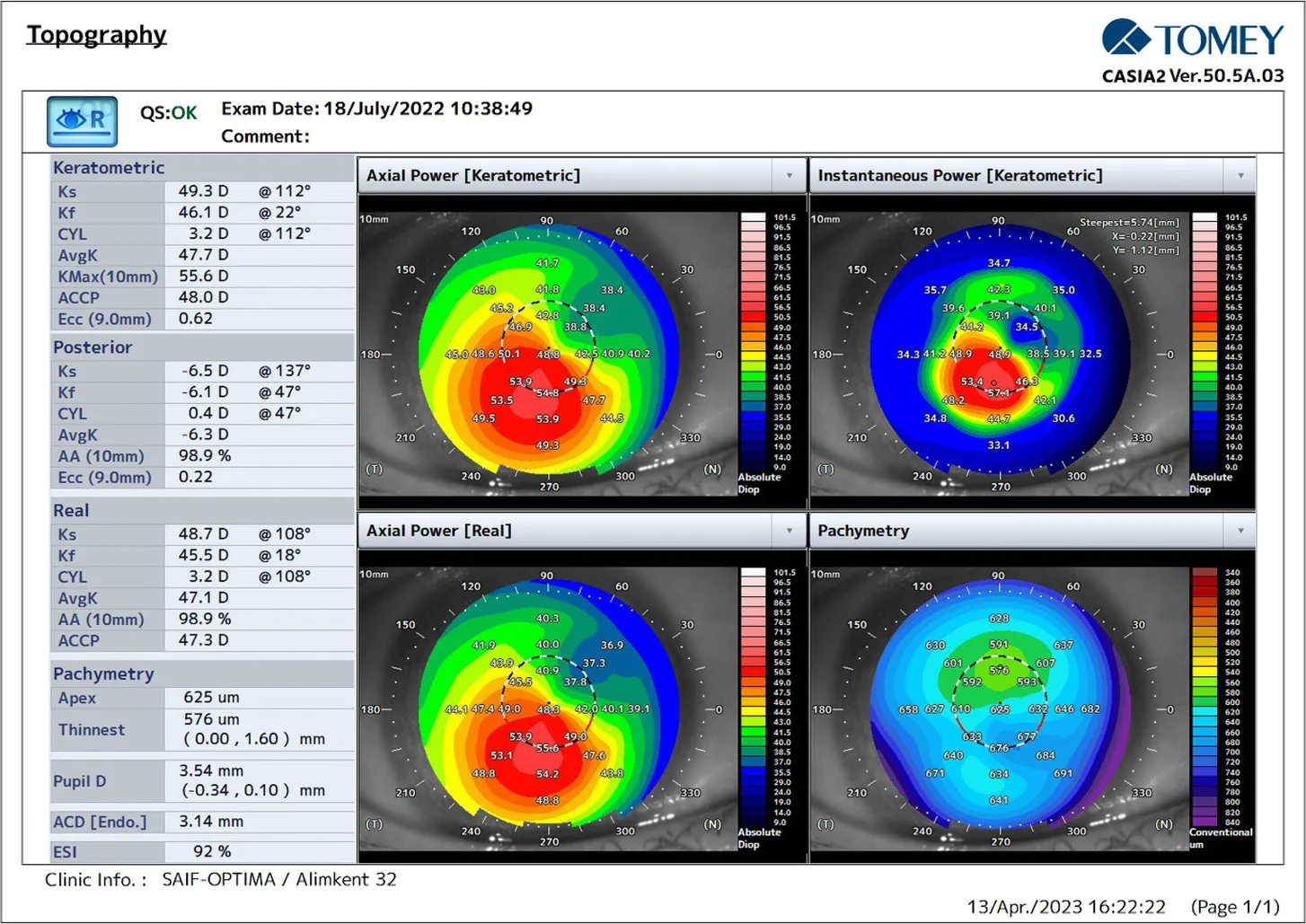
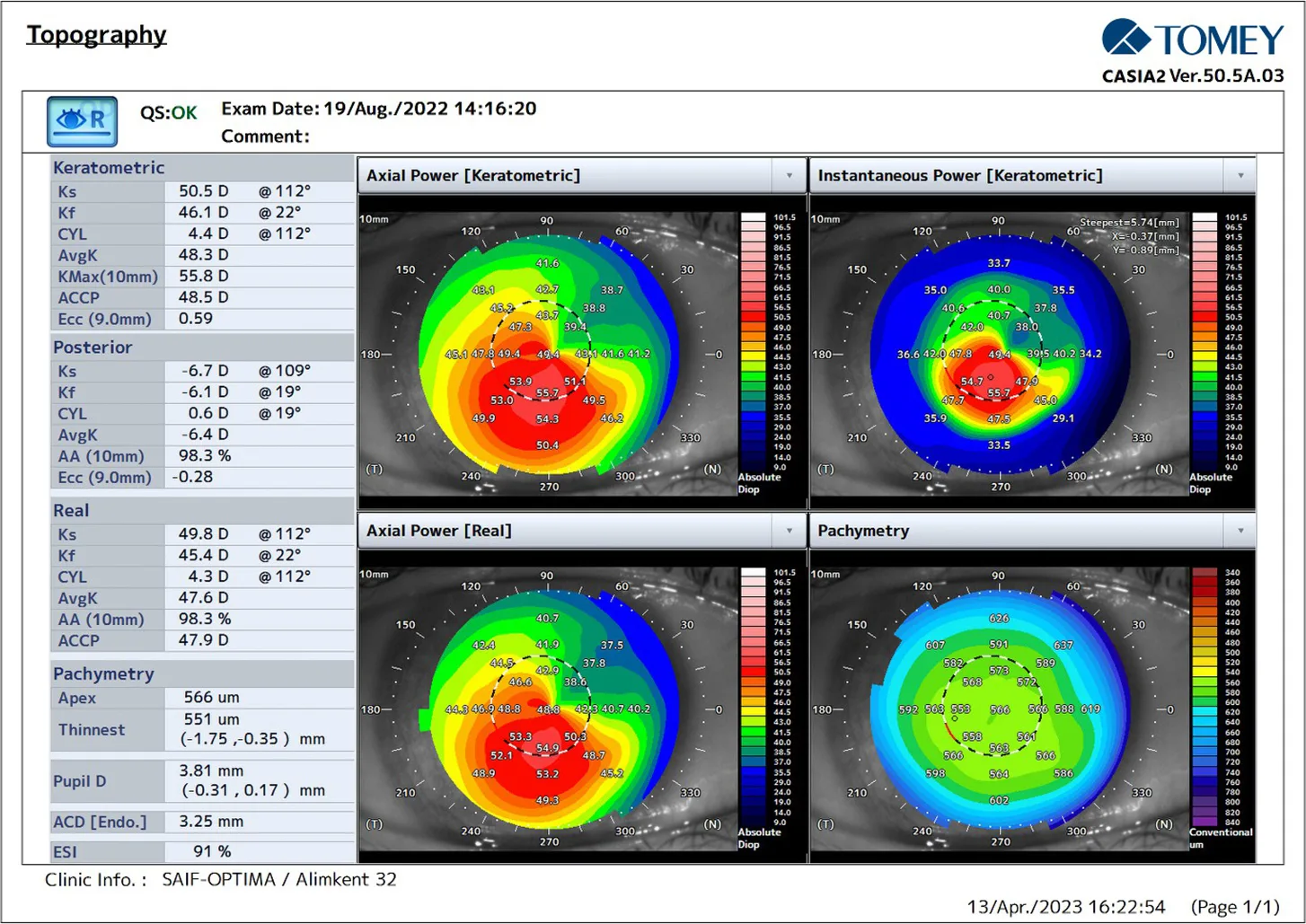
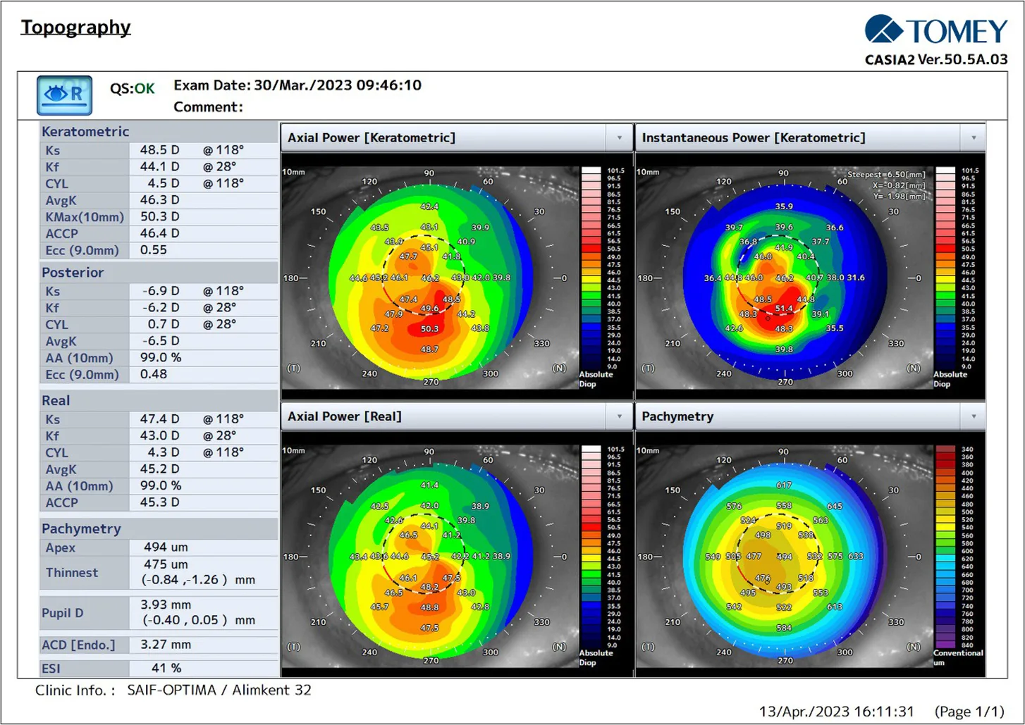
| 2 Months | Stable morphology, VA 0.3 | No complications |
| 9 Months | VA 0.5–0.8, normal topography | Long-term stabilisation |
Further Questions?
Do you have any questions or require further information?
We would be pleased if you send us an inquiry.