
Advanced Imaging of Preserflo Microshunt in Uveitic Glaucoma Treatment
This is a 50-year-old male patient who had a history of uveitic glaucoma in his left eye, with previous Non-Penetrating Deep Sclerectomy surgery. It was decided to re-intervene in this eye after presenting pressures of 30 mmHg, performing a Preserflo Microshunt surgery (Santen, Osaka, Japan) in the superior temporal quadrant.
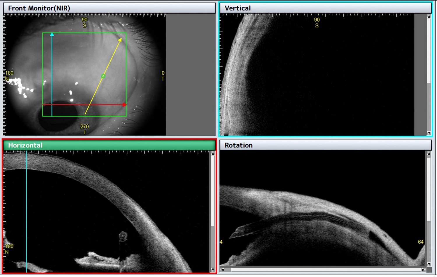
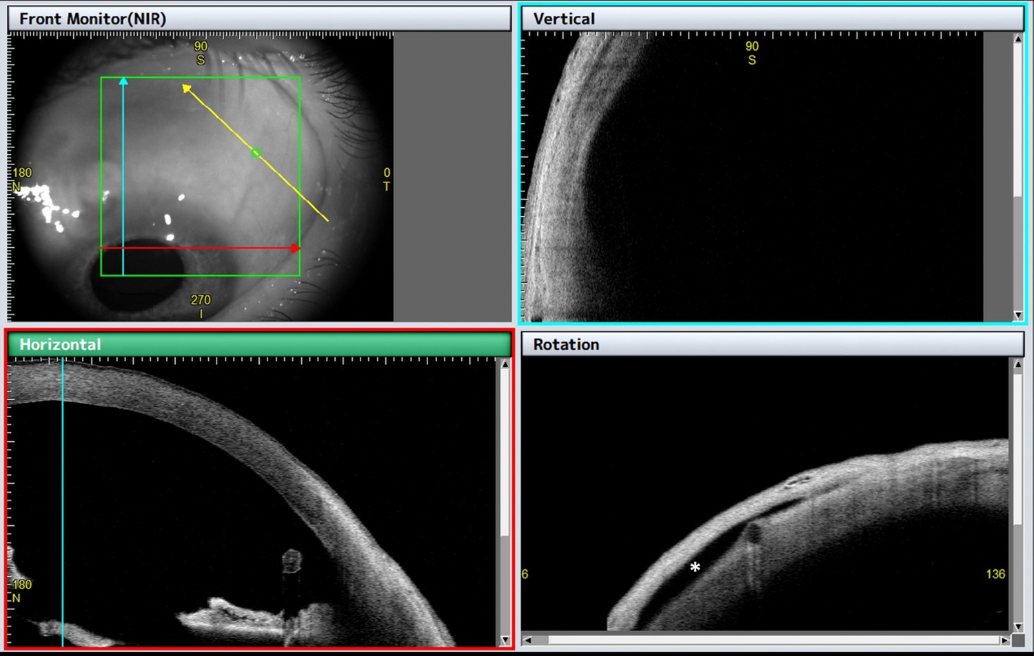
CASIA2 images allow us to locate the intraocular portion of the implant, calculate the distance from the tip of the tube to endothelium, as well as visualise the entire tube pathway, its internal lumen (figure 1), and the presence of posterior conjunctival bleb, visualised as a hyporeflective area in the most posterior part of the implant (asterisk in figure 2).
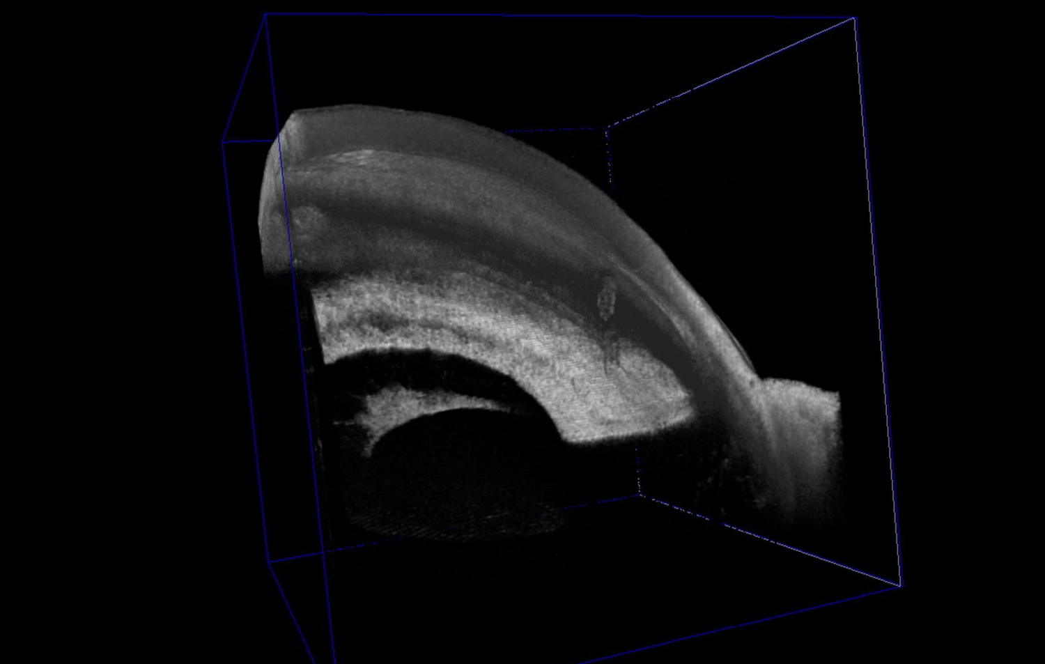
We can also make a 3D reconstruction of anterior segment localising the tube entrance (figure 3). The current pressure is 10 mmHg.
Further Questions?
Do you have any questions or require further information?
We would be pleased if you send us an inquiry.